International CME for Today's Radiologist
Global Radiology CME
Search Results
256 results found with an empty search
- Brachydactyly Type A3
50-year-old female presenting with short 5th finger . What is the diagnosis? • Xray of the Week Figure 1. Frontal bilateral hand X-ray. What is the diagnosis? Figure 2. Frontal bilateral hand X-ray: Severe shortening of the middle phalanx of the fifth digit (red arrows). The phalanx measures less than 50% of the length of the adjacent fourth middle phalanx, satisfying the Hertzog criteria for Brachydactyly Type A3. A mild radial clinodactyly is present, secondary to the wedge-shaped morphology of the middle phalanx. Discussion Brachydactyly (BD) refers to disproportionately short fingers and toes, classified by the Julia Bell system into five primary types (A–E) based on anatomical involvement. Type A3 (BDA3), also known as brachymesophalangy V, is the most common isolated hand anomaly.[1] Imaging Findings and Analysis Radiographs of this 50-year-old female (Fig 1, 2) reveal isolated, bilateral shortening of the middle phalanx of the fifth digit. The definitive diagnosis is established using the Hertzog criterion, which mandates that the longitudinal length of the fifth middle phalanx must be less than 50% of the longitudinal length of the fourth middle phalanx.[1] Morphologically, the affected phalanx exhibits a characteristic rhomboid or wedge-shaped configuration. The radial side of the phalanx is significantly shorter than the ulnar side, creating a slanted distal articular surface that drives radial clinodactyly (radial deflection of the distal phalanx toward the fourth digit). Pathophysiological and Genetic Mechanisms BDA3 is an autosomal dominant disorder arising from disrupted cartilage ossification during early embryonic development (blastogenesis), with some cases mapped to the chromosome 13q33 region.[1] Epidemiology and Biocultural Insights The prevalence of BDA3 varies significantly by ethnicity; it is a common variant in Asian populations, occurring in 21% to 25.6% of Japanese children, but remains rare in populations of European or African descent (<2%).[7][8] Growth and Development Although BDA3 is more frequently identified in children with short stature, it does not negatively impact the efficacy of growth hormone therapy, indicating that the local physis disruption does not represent global resistance to growth signals.[4] Syndromic Associations and Differentials Radiologists should view BDA3 as a clinical marker for broader genetic conditions, most notably Down syndrome (present in ~60%) and Turner syndrome.[9] Differential considerations include Kirner deformity (palmo-radial bowing of the distal phalanx shaft) and Camptodactyly (a soft-tissue flexion contracture of the PIP joint).[5] Genetic Counseling Inheritance is typically autosomal dominant with a 50% recurrence risk for offspring; however, for isolated cases, patients should be reassured that BDA3 is a benign anatomical variant.[1] Management and Prognosis Isolated BDA3 is almost universally asymptomatic and requires no medical or surgical intervention. The functional prognosis is excellent, as the shortening of the little finger does not typically affect manual dexterity or grip strength.[5] Corrective surgical procedures, such as osteotomy, are extremely rare and indicated only for severe clinodactyly that causes functional impairment or significant cosmetic distress.[1] Key Learning Points Diagnostic Standard : BDA3 is confirmed when the fifth middle phalanx length is <50% of the fourth (Hertzog criterion) .[1] Radiographic Features : Look for a wedge-shaped middle phalanx and associated radial clinodactyly. Population Variant : High prevalence in Japanese and other Asian populations should be noted as a common anatomical variation .[7][8] Clinical Marker : Identification of BDA3 warrants screening for Down and Turner syndromes, particularly if associated with metacarpal shortening .[9] Management : Isolated BDA3 is benign; functional impairment is rare, though corrective options exist for severe cases .[5] References Temtamy SA, Aglan MS. Brachydactyly. Orphanet J Rare Dis . 2008;3:15. doi: 10.1186/1750-1172-3-15 Garn SM, Hertzog KP, Poznanski AK, Nagy JM. Metacarpophalangeal length in the evaluation of skeletal malformation. Radiology . 1972;105(2):375-381. doi: 10.1148/105.2.375 Wu H, Wu H, Li Y, Li H. Brachydactyly Type A3 Is More Commonly Seen in Children With Short Stature But Does Not Affect Their Height Improvement by Growth Hormone Therapy. Front Endocrinol (Lausanne) . 2022;13:824315. doi: 10.3389/fendo.2022.824315 Nguyen ML, Jones N. Undergrowth: brachydactyly. Hand Clin . 2009;25(2):247-255. doi: 10.1016/j.hcl.2009.02.003 Everman DB. The brachydactylies. In: Stevenson RE, Hall JG, eds. Human Malformations and Related Anomalies . 2nd ed. Oxford University Press; 2006:968-983. Zhang W, Li K, Zhang Q, et al. Epidemiology of brachydactyly type A3 in China: a nationwide multicentre population-based study among children aged 3–17 years. BMJ Open . 2025;15(11):e099166. doi: 10.1136/bmjopen-2025-099166 Wu HH, Zhang YQ, Yu CD, et al. Brachydactyly type A3 may be associated with shorter stature: An observation from a Chinese pediatric sample. PLoS One . 2025;20(11):e0336913. doi: 10.1371/journal.pone.0336913 Kang MJ, Kanakatti Shankar R, Jee YH. Phalangeal bone growth and implications in Turner syndrome. Front Endocrinol (Lausanne) . 2026;16:1735962. doi: 10.3389/fendo.2025.1735962 . Castriota-Scanderbeg A, Dallapiccola B. Abnormal Skeletal Phenotypes: From Sameness to Differential Diagnosis . Springer; 2006. doi: 10.1007/3-540-30361-8 Cleveland Clinic. Brachydactyly. Accessed January 20, 2026. https://my.clevelandclinic.org/health/diseases/24081-brachydactyly Kevin M. Rice, MD is the president of Global Radiology CME and is a radiologist with Cape Radiology Group . He has held several leadership positions including Board Member and Chief of Staff at Valley Presbyterian Hospital in Los Angeles, California. Dr. Rice has made several media appearances as part of his ongoing commitment to public education. Dr. Rice's passion for state-of-the-art radiology and teaching includes acting as a guest lecturer at UCLA. In 2015, Dr. Rice and Natalie Rice founded Global Radiology CME to provide innovative radiology education at exciting international destinations, with the world's foremost authorities in their field. In 2016, Dr. Rice was nominated and became a semifinalist for a "Minnie" Award for the Most Effective Radiology Educator. He was once again a semifinalist for a "Minnie" for 2021's Most Effective Radiology Educator by AuntMinnie.com . He has continued to teach by mentoring medical students interested in radiology . Everyone he has mentored has been accepted into top programs across the country, including Harvard, UC San Diego, Northwestern, Vanderbilt, and Thomas Jefferson. Follow Dr. Rice on Twitter @KevinRiceMD All posts by Kevin M. Rice, MD
- Atrial Lead Dislodgement in Dual-Chamber Permanent Pacemaker
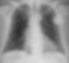
An 88-year-old male status post dual-lead permanent pacemaker placement presented with symptoms of pacemaker malfunction, including bradycardia and fatigue. What is the diagnosis? • Xray of the Week Figure 1. Frontal chest X-ray. What is the diagnosis? Figure 2. Frontal chest X-ray: Both pacemaker leads project over the right ventricle (red and green arrows), with the intended atrial lead dislodged from the right atrium and positioned in the right ventricle alongside the ventricular lead. The pulse generator is appropriately located in the left pectoral region. Diagnosis Atrial lead dislodgement in dual-lead permanent pacemaker, with both leads in the right ventricle. Discussion Pacemaker lead dislodgement is a common early complication of dual-lead permanent pacemaker implantation, occurring in 1-5% of cases, often due to inadequate fixation, patient activity, or twiddler's syndrome (patient manipulation of the device).[1][2] In dual-lead systems , the atrial lead is typically placed in the right atrial appendage for sensing and pacing, while the ventricular lead is in the right ventricular apex. Dislodgement of the atrial lead into the right ventricle can lead to ineffective atrial pacing, loss of atrioventricular synchrony, and symptoms of pacemaker syndrome or arrhythmia.[3][4] Radiologically, this manifests as abnormal lead trajectory on chest X-ray, potentially causing complications like perforation, thrombosis, or tricuspid valve injury if unrecognized.[5] Imaging Findings Chest X-ray is the first-line modality for assessing pacemaker lead position and integrity post-implantation, with high sensitivity for detecting dislodgement (nearly 100% when compared to electrocardiography).[1][6] Frontal and lateral views are essential; lateral views help confirm chamber placement, as frontal views alone may be ambiguous due to overlap. On frontal chest X-ray: The normal atrial lead shows a gentle J-curve toward the right atrium (superior and lateral), while the ventricular lead has a straighter course to the right ventricular apex (inferior and medial). In dislodgement, the atrial lead deviates inferiorly, projecting over the right ventricle similar to the ventricular lead, often appearing parallel or crossed. Associated findings: Lead redundancy, coiling, or fracture; pulse generator rotation (in twiddler's syndrome ); or signs of perforation (e.g., lead tip beyond cardiac silhouette). Multiplanar CT can be used for confirmation if X-ray is equivocal, but routine post-implantation chest X-ray is sufficient for most cases.[2][7] Lead Type Normal Position on Frontal Chest X-ray Abnormal (Dislodged) Position Atrial Lead J-shaped curve directing superior-laterally to right atrial appendage Straight or inferior course to right ventricle, parallel to ventricular lead Ventricular Lead Straight course to right ventricular apex, tip pointing inferior-medially May be unaffected, but both leads in RV if atrial dislodged Management and Prognosis Management involves urgent lead repositioning via percutaneous extraction and reimplantation, with electrocardiographic confirmation of capture; anticoagulation may be needed if thrombosis is suspected.[3][5] Prognosis is excellent with early detection (success rate >95%), but delayed recognition can lead to reoperation or complications like heart failure (mortality <1% in uncomplicated cases).[4][6] Key Learning Points Chest X-ray is critical for immediate post-implantation evaluation of pacemaker lead position, identifying dislodgement by abnormal trajectory. Look for lack of J-curve in the atrial lead and both leads projecting over the right ventricle on frontal views; obtain lateral views to confirm chamber. Radiology interpretation guides timely intervention, preventing complications like perforation or loss of pacing—always compare to prior films. Routine chest X-ray post-procedure detects most lead-related issues; correlate with ECG for functional assessment. References Costelloe CM. Radiography of pacemakers and implantable cardioverter defibrillators. AJR Am J Roentgenol . 2012;199(6):1252-1258. doi: 10.2214/AJR.12.8641 Silva C, Christensen JD. How I do it: evaluating cardiac implantable devices and noncardiac mimics on chest radiographs. Radiology . 2025;315(2):e241911. doi: 10.1148/radiol.241911 Hunter TB, Taljanovic MS, Tsau PH, Berger WG, Standen JR. Medical devices of the chest. Radiographics . 2004;24(6):1725-1746. doi: 10.1148/rg.246045031 Aguilera AL, Volokhina YV, Fisher KL. Radiography of cardiac conduction devices: a comprehensive review. Radiographics . 2011;31(6):1669-1682. doi: 10.1148/rg.316115529 Dipoce J, Soni A, Dhillon S, Golzarian J. Radiology of cardiac devices and their complications. Br J Radiol . 2015;88(1046):20140540. doi: 10.1259/bjr.20140540 Pascual Alandete Germán S, Isarria Vidal S, Domingo Montañana ML, De la Vía Oraá E, Vilar Samper J. Pacemakers and implantable cardioverter defibrillators, unknown to chest radiography: review, complications and systematic reading. Eur J Radiol . 2015;84(3):499-508. doi: 10.1016/j.ejrad.2014.12.011 Mathew RP, Alexander T, Patel V, Low G. Chest radiographs of cardiac devices (Part 1): cardiovascular implantable electronic devices, cardiac valve prostheses and Amplatzer occluder devices. SA J Radiol . 2019;23(1):1730. doi: 10.4102/sajr.v23i1.1730 Kevin M. Rice, MD is the president of Global Radiology CME and is a radiologist with Cape Radiology Group . He has held several leadership positions including Board Member and Chief of Staff at Valley Presbyterian Hospital in Los Angeles, California. Dr. Rice has made several media appearances as part of his ongoing commitment to public education. Dr. Rice's passion for state-of-the-art radiology and teaching includes acting as a guest lecturer at UCLA. In 2015, Dr. Rice and Natalie Rice founded Global Radiology CME to provide innovative radiology education at exciting international destinations, with the world's foremost authorities in their field. In 2016, Dr. Rice was nominated and became a semifinalist for a "Minnie" Award for the Most Effective Radiology Educator. He was once again a semifinalist for a "Minnie" for 2021's Most Effective Radiology Educator by AuntMinnie.com . He has continued to teach by mentoring medical students interested in radiology . Everyone he has mentored has been accepted into top programs across the country, including Harvard, UC San Diego, Northwestern, Vanderbilt, and Thomas Jefferson. Follow Dr. Rice on Twitter @KevinRiceMD All posts by Kevin M. Rice, MD
- Arachnoiditis Ossificans
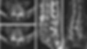
An 74-year-old male with chronic low back pain and progressive lower extremity weakness. What is the diagnosis? • Xray of the Week Figure 1. Non-contrast CT of the lumbar spine. What is the diagnosis? Figure 2. Non-contrast CT of the lumbar spine (axial, sagittal, and coronal reconstructions): Central pattern of arachnoiditis ossificans demonstrating dagger-like ossification within the central spinal canal at the L5 level (red arrows). The linear, hyperdense bony bar courses longitudinally along the central canal, causing focal narrowing and potential cauda equina compression. No peripheral or circumferential involvement is seen, consistent with the central ossification pattern. Diagnosis Arachnoiditis ossificans. Discussion Arachnoiditis ossificans is a rare end-stage manifestation of chronic adhesive arachnoiditis, characterized by ossification of the arachnoid membrane within the spinal canal.[1][2] It typically occurs in the thoracolumbar region and is associated with prior spinal surgery, trauma, infection, subarachnoid hemorrhage, or oil-based myelography.[3][4] Patients often present with progressive myelopathy or radiculopathy due to nerve root compression or tethering.[5] The condition represents metaplastic ossification of inflamed arachnoid tissue, leading to intrathecal calcified or ossified plaques that can encase the spinal cord or cauda equina.[6] Imaging Findings Computed tomography (CT) is the modality of choice for diagnosing arachnoiditis ossificans due to its superior depiction of ossified structures, with sensitivity for detecting intrathecal ossification approaching 100% in symptomatic cases.[2][7] On CT, hyperdense bone attenuation plaques are seen within the thecal sac, with patterns varying by type of ossification [2]: Central pattern : Linear or dagger-like hyperdense ossification centered within the spinal canal, often appearing as a solitary midline bony bar or rod on axial views. (Figs. 1,2) Nerve root encasing pattern : Tubular or circumferential hyperdense ossifications surrounding individual or clumped nerve roots (cauda equina), with roots appearing embedded or passing through the ossified tissue. Weblike pattern : Interlacing, mesh-like hyperdense strands or trabeculae filling or crisscrossing the thecal sac, creating a reticular appearance between nerve roots. Peripheral pattern : Rim-like or discontinuous hyperdense plaques along the inner walls of the thecal sac, often circumferential or partially encircling, potentially narrowing the canal without central involvement. These patterns frequently coexist, leading to thecal sac deformity, nerve root clumping, and varying degrees of central canal stenosis. Multiplanar reconstructions (sagittal/coronal) help confirm the distribution and extent. Patterns of intrathecal ossification can guide prognosis and surgical planning. Management and Prognosis Management is primarily conservative, focusing on pain control and physical therapy, as surgical decompression carries high risks of reossification or worsening symptoms.[3][5] Laminectomy with duraplasty may be considered for severe cord compression, with variable success rates (30-50% improvement in symptoms).[4] Prognosis is guarded, with many patients experiencing progressive disability; early detection via imaging can facilitate timely intervention to prevent irreversible myelopathy.[6] Key Learning Points CT is essential for identifying hyperdense intrathecal ossifications in arachnoiditis ossificans, outperforming MRI for bony detail. Look for circumferential or patterned ossified plaques encasing nerve roots on multiplanar reconstructions to differentiate from mimics like dural calcifications. Radiology plays a critical role in classifying ossification patterns, which influence surgical feasibility and prognosis. Prompt recognition on CT can guide multidisciplinary management and prevent complications like syringomyelia—always correlate with clinical history of prior spinal insult. References Frizzell B, Kaplan P, Dussault R, Sevick R. Arachnoiditis ossificans: MR imaging features in five patients. AJR Am J Roentgenol . 2001;177(2):461-464. doi: 10.2214/ajr.177.2.1770461 Thejeel B, Greditzer-Sobeck C, Ciacci J, Siddiqi I. Patterns of intrathecal ossification in arachnoiditis ossificans: a retrospective case series. AJNR Am J Neuroradiol . 2023;44(2):228-234. doi: 10.3174/ajnr.A7764 Donalisio M, Cadosch D. Arachnoiditis ossificans. Skeletal Radiol . 2024;53(5):1019-1021. doi: 10.1007/s00256-023-04465-7 Junewick JJ. Clinical image. Arachnoiditis ossificans in a pediatric patient. Pediatr Radiol . 2010;40(2):228. doi: 10.1007/s00247-009-1350-2 Jaspan T, Preston BJ, Mulholland RC, Webb JK. The CT appearances of arachnoiditis ossificans. Spine (Phila Pa 1976) . 1990;15(2):148-151. doi: 10.1097/00007632-199002000-00022 Chan CC, Lau PY, Sun LK, Lo SS. Arachnoiditis ossificans. Hong Kong Med J. 2009;15(2):146-148. https://pubmed.ncbi.nlm.nih.gov/19342743/ Kumaran SP, Gupta K, Maddali A, Viswamitra S. Post traumatic arachnoiditis ossificans. J Emerg Trauma Shock . 2012;5(3):250-252. doi: 10.4103/0974-2700.99701 Sefczek RJ, Deeb ZL. Case report: computed tomography findings in spinal arachnoiditis ossificans. J Comput Tomogr . 1983;7(3):315-318. doi: 10.1016/0149-936x(83)90099-1 Kevin M. Rice, MD is the president of Global Radiology CME and is a radiologist with Cape Radiology Group . He has held several leadership positions including Board Member and Chief of Staff at Valley Presbyterian Hospital in Los Angeles, California. Dr. Rice has made several media appearances as part of his ongoing commitment to public education. Dr. Rice's passion for state-of-the-art radiology and teaching includes acting as a guest lecturer at UCLA. In 2015, Dr. Rice and Natalie Rice founded Global Radiology CME to provide innovative radiology education at exciting international destinations, with the world's foremost authorities in their field. In 2016, Dr. Rice was nominated and became a semifinalist for a "Minnie" Award for the Most Effective Radiology Educator. He was once again a semifinalist for a "Minnie" for 2021's Most Effective Radiology Educator by AuntMinnie.com . He has continued to teach by mentoring medical students interested in radiology . Everyone he has mentored has been accepted into top programs across the country, including Harvard, UC San Diego, Northwestern, Vanderbilt, and Thomas Jefferson. Follow Dr. Rice on Twitter @KevinRiceMD All posts by Kevin M. Rice, MD
- Hyphema with Vitreous Hemorrhage
Pt fell and his eye hit a metal object. What is the diagnosis? • Xray of the Week Figure 1. Non-contrast CT orbits – axial and sagittal images. What is the diagnosis? Figure 2. Non-contrast CT orbits – axial and sagittal images. Hyperdense material consistent with blood fills the anterior chamber of the left eye (red arrows), indicating hyphema. Hyperdense material is also present in the vitreous chamber posteriorly (yellow arrows), consistent with acute vitreous hemorrhage. The right globe shows a normal lens separating anterior and posterior chambers (green arrows) for comparison. Diagnosis Traumatic hyphema with concurrent vitreous hemorrhage in the left eye. Discussion Hyphema is hemorrhage into the anterior chamber, most commonly from blunt or penetrating ocular trauma, but also possible from surgery, coagulopathy, or spontaneous causes (e.g., iris neovascularization).[1][2] Vitreous hemorrhage involves bleeding into the vitreous cavity, often linked to trauma, proliferative diabetic retinopathy, retinal tears, or vascular occlusions.[3] In this traumatic case, both findings coexist due to shearing forces disrupting iris/ciliary body vessels (hyphema) and posterior structures (vitreous hemorrhage). Hyphemas are graded clinically by the volume of layered blood in the anterior chamber (Table below). Grading helps predict complications like rebleeding (highest in grades III–IV) and secondary glaucoma.[4][5][6] Hyphema Grade Approximate Volume of Blood in the Anterior Chamber Risk of IOP elevation/ secondary glaucoma Grade 0: Microhyphema <1% (slit-lamp only) <10% Grade I <33% ~10% Grade II 33–50% ~10% Grade III >50% but not full ~25% Grade IV 100% >50% "Eight ball" hyphema 100% & dark color due to poor circulation 100% Figure 3. Traumatic hyphema c linical image: Note the layering blood in the anterior chamber in this patient following blunt eye trauma. Contributor : Jesse Vislisel, MD - EyeRounds.org The University of Iowa. Creative Commons 3.0 https://webeye.ophth.uiowa.edu/eyeforum/atlas/pages/Hyphema/index.htm#gsc.tab=0 Imaging Findings Non-contrast orbital CT is the preferred initial imaging for acute ocular trauma when direct exam is limited by swelling, pain, or suspected open globe.[7][8] Hyphema appears as hyperdense (blood attenuation ~40–70 HU) material layering or filling the anterior chamber, anterior to the lens.[7] Vitreous hemorrhage shows increased attenuation in the posterior segment, often homogeneous acutely or heterogeneous if clotted/organized.[3][9] Reported sensitivity for detecting hyphema on CT is ~77%, specificity ~88%.[7] CT excels at identifying associated injuries (e.g., lens dislocation, globe rupture, foreign bodies, orbital fractures) and is safer than ultrasound if open globe is suspected (to avoid pressure on the eye).[8] Ultrasound is superior for posterior segment details (e.g., retinal detachment ) when media opacity from hemorrhage limits fundus view, but CT is first-line for trauma screening.[3][9] Management & Prognosis Management is primarily ophthalmology-directed. For traumatic hyphema: conservative measures include protective eye shielding, head elevation (30–45°), limited activity/bed rest, avoiding aspirin/NSAIDs/anticoagulants, and serial IOP monitoring to prevent rebleeding (peak risk days 3–5) or secondary glaucoma.[4][5][6][10] Topical corticosteroids reduce inflammation; cycloplegics (e.g., atropine) relieve ciliary spasm/pain. Antifibrinolytics (e.g., tranexamic acid or aminocaproic acid) may reduce rebleeding risk but lack strong evidence for improving final visual acuity and can prolong clot clearance—use is controversial per recent reviews.[10] There are ongoing debates regarding optimal approaches due to the absence of standardized guidelines, including medical agents, surgical techniques, and special situations (e.g., sickle-cell disease).[8] Surgical evacuation (e.g., anterior chamber washout) is indicated for large/persistent hyphema causing corneal blood staining, uncontrolled IOP, or active rebleeding. Vitreous hemorrhage often resolves spontaneously but may require vitrectomy if non-clearing or associated with retinal pathology.[3] Prognosis is generally good for isolated/low-grade cases (most resolve within days to weeks), but worse with higher grades, rebleeding, secondary glaucoma, sickle cell trait/disease, or extensive posterior involvement (e.g., retinal detachment). Approximately 5% of traumatic hyphemas require surgery.[4][5] Key Learning Points Look for hyperdense anterior chamber fluid on non-contrast CT to diagnose hyphema in trauma. Vitreous hyperdensity indicates posterior hemorrhage; always assess for associated globe/orbital injuries. CT is valuable when clinical exam is limited; ultrasound complements for posterior evaluation. Grade hyphema clinically to guide prognosis and intervention—prompt ophthalmology consultation is essential to prevent vision loss. References Sung EK, Nadgir RN, Fujita A, et al. Injuries of the globe: what can the radiologist offer? Radiographics . 2014;34(3):764-776. doi: 10.1148/rg.343135120 Hallinan JTPD, Pillay P, Koh L, Goh K, Yu W. Eye globe abnormalities on MR and CT in adults: an anatomical approach. Korean J Radiol . 2016;17(5):664-673. doi: 10.3348/kjr.2016.17.5.664 Spraul CW, Grossniklaus HE. Vitreous hemorrhage. Surv Ophthalmol . 1997;42(1):3-39. doi: 10.1016/S0039-6257(97)84041-6 Hartness E, Garza Reyes A, Yu C, Sears N. Hyphema: diagnosis and management. EyeRounds.org . February 20, 2024. Accessed January 14, 2026. https://eyerounds.org/cases/345-hyphema.htm Chen EJ, Fasiuddin A. Management of traumatic hyphema and prevention of its complications. Cureus . 2021;13(6):e15771. doi: 10.7759/cureus.15771 Miller SC, Meeralakshmi P, Fliotsos MJ, et al. Global current practice patterns for the management of hyphema. Clin Ophthalmol . 2022;16:3135-3144. doi: 10.2147/OPTH.S372273 Chazen JL, El-Sayed IH, Vance S, et al. CT in the evaluation of acute injuries of the anterior eye segment. AJR Am J Roentgenol . 2018;210(3):W107-W113. doi: 10.2214/AJR.17.18279 Bansal S, Gunasekaran PK, Azad S, Agrawal R. Controversies in the pathophysiology and management of hyphema. Surv Ophthalmol . 2016;61(3):297-308. doi: 10.1016/j.survophthal.2015.11.005 Rabinowitz R, Yagev R, Shoham A, Lifshitz T. Comparison between clinical and ultrasound findings in patients with vitreous hemorrhage. Eye (Lond) . 2004;18(3):253-256. doi: 10.1038/sj.eye.6700632 Woreta FA, Lindsley KB, Gharaibeh A, et al. Medical interventions for traumatic hyphema. Cochrane Database Syst Rev . 2023;2023(3):CD005431. doi: 10.1002/14651858.CD005431.pub5 Kevin M. Rice, MD is the president of Global Radiology CME and is a radiologist with Cape Radiology Group . He has held several leadership positions including Board Member and Chief of Staff at Valley Presbyterian Hospital in Los Angeles, California. Dr. Rice has made several media appearances as part of his ongoing commitment to public education. Dr. Rice's passion for state of the art radiology and teaching includes acting as a guest lecturer at UCLA. In 2015, Dr. Rice and Natalie Rice founded Global Radiology CME to provide innovative radiology education at exciting international destinations, with the world's foremost authorities in their field. In 2016, Dr. Rice was nominated and became a semifinalist for a "Minnie" Award for the Most Effective Radiology Educator. He was once again a semifinalist for a "Minnie" for 2021's Most Effective Radiology Educator by AuntMinnie.com . He has continued to teach by mentoring medical students interested in radiology . Everyone who he has mentored has been accepted into top programs across the country including Harvard, UC San Diego, Northwestern, Vanderbilt, and Thomas Jefferson. Follow Dr. Rice on Twitter @KevinRiceMD All posts by Kevin M. Rice, MD
- Active Bleeding in the Ascending Colon
An 83-year-old male with rectal bleeding. What is the diagnosis? • Xray of the Week Figure 1. CTA abdomen/pelvis. What is the diagnosis? Figure 2. CT angiogram: A. Non-contrast images are normal (yellow arrow) . B. Arterial phase image: Hyperdense extravasation of contrast within the lumen of the ascending colon (blue arrow), indicating active bleeding. C and D. Note the dependent layering of contrast on portal venous phase images, confirming active extravasation. Diagnosis Active gastrointestinal bleeding in the ascending colon. Discussion Gastrointestinal (GI) bleeding is a common clinical problem, particularly in the elderly, where lower GI sources predominate. Common etiologies include diverticulosis, angiodysplasia, neoplasms, ischemia, and inflammatory conditions.[1][2] In this case, the ascending colon involvement suggests a right-sided colonic source, often angiodysplasia or diverticular bleed, which can present with painless hematochezia.[3] Active bleeding is defined as extravasation of contrast into the bowel lumen on imaging, distinguishing it from pseudo-extravasation mimics like hyperdense pills or fecal material.[4] Imaging Findings Multiphase computed tomography angiography (CTA) is the preferred initial imaging for hemodynamically stable patients with suspected acute GI bleeding, offering high sensitivity (85-89%) and specificity (85-95%) for detecting active extravasation.[5][6] It is rapid, widely available, and guides subsequent interventions like endoscopy or embolization.[7] On non-contrast phase: No extravasation; may show hyperdense sentinel clot (40-70 HU) or bowel wall abnormalities. On arterial phase: Active extravasation appears as hyperdense focus (>90 HU) within the lumen, often eccentric or jet-like, increasing in size or density compared to non-contrast. On portal venous/delayed phase: Extravasation persists or enlarges, confirming active bleed; helps differentiate from venous sources. Reported detection rates for active bleeding exceed 0.3-0.5 mL/min, superior to nuclear medicine in many settings.[8] CTA also identifies structural causes (e.g., diverticula, tumors) even without active bleeding.[4] Management and Prognosis Management begins with hemodynamic resuscitation. For active lower GI bleeding confirmed on CTA, options include urgent colonoscopy (if stable), interventional radiology embolization (targeted to the bleeding vessel), or surgery for refractory cases.[1][3][7] Embolization success rates are 70-90%, with low rebleeding if superselective.[2] Prognosis depends on bleed severity, comorbidities, and etiology; elderly patients have higher mortality (10-20% for severe bleeds). Rebleeding risk is 10-25% for diverticular sources.[6] Key Learning Points Multiphase CTA is first-line for localizing active GI bleeding in stable patients, with high accuracy for extravasation. Look for hyperdense intraluminal contrast on post-contrast phases that is absent on non-contrast to confirm active bleed. CTA guides therapy by identifying the site and potential etiology; always assess for mimics like hyperdense foreign material. Prompt radiology interpretation is crucial to expedite embolization or endoscopy and improve outcomes. References Artigas JM, Martí M, Soto JA, Esteban H, Pinilla I, Guillén E. Multidetector CT angiography for acute gastrointestinal bleeding: technique and findings. Radiographics . 2013;33(5):1453-1470. doi: 10.1148/rg.335125072 Wells ML, Hansel SL, Bruining DH, et al. CT for evaluation of acute gastrointestinal bleeding. Radiographics . 2018;38(4):1089-1107. doi: 10.1148/rg.2018170138 Wortman JR, Landman W, Fulwadhva UP, Viscomi SG, Sodickson AD. CT angiography for acute gastrointestinal bleeding: what the radiologist needs to know. Br J Radiol . 2017;90(1075):20170076. doi: 10.1259/bjr.20170076 Di Serafino M, Iacobellis F, Schillirò ML, et al. The role of CT-angiography in the acute gastrointestinal bleeding: a pictorial essay of active and obscure findings. Tomography . 2022;8(5):2369-2402. doi: 10.3390/tomography8050198 Wu LM, Xu JR, Yin Y, Qu XH. Usefulness of CT angiography in diagnosing acute gastrointestinal bleeding: a meta-analysis. World J Gastroenterol . 2010;16(31):3957-3963. doi: 10.3748/wjg.v16.i31.3957 García-Blázquez V, Vicente-Bártulos A, Olavarria-Delgado A, Plana MN, van der Winden D, Zamora J. Accuracy of CT angiography in the diagnosis of acute gastrointestinal bleeding: systematic review and meta-analysis. Eur Radiol . 2013;23(5):1181-1190. doi: 10.1007/s00330-012-2721-x Kim BS, Li BT, Engel A, Samra JS, Clarke S, Norton ID, Li AE. Diagnosis of gastrointestinal bleeding: a practical guide for clinicians. World J Gastrointest Pathophysiol . 2014;5(4):467-478. doi: 10.4291/wjgp.v5.i4.467 Parekh PJ, Buerlein RC, Shams R, Vingan H, Johnson DA. Evaluation of gastrointestinal bleeding: update of current radiologic strategies. World J Gastrointest Pharmacol Ther. 2014;5(4):200-208. doi: 10.4292/wjgpt.v5.i4.200 Kevin M. Rice, MD is the president of Global Radiology CME and is a radiologist with Cape Radiology Group . He has held several leadership positions including Board Member and Chief of Staff at Valley Presbyterian Hospital in Los Angeles, California. Dr. Rice has made several media appearances as part of his ongoing commitment to public education. Dr. Rice's passion for state-of-the-art radiology and teaching includes acting as a guest lecturer at UCLA. In 2015, Dr. Rice and Natalie Rice founded Global Radiology CME to provide innovative radiology education at exciting international destinations, with the world's foremost authorities in their field. In 2016, Dr. Rice was nominated and became a semifinalist for a "Minnie" Award for the Most Effective Radiology Educator. He was once again a semifinalist for a "Minnie" for 2021's Most Effective Radiology Educator by AuntMinnie.com . He has continued to teach by mentoring medical students interested in radiology . Everyone he has mentored has been accepted into top programs across the country, including Harvard, UC San Diego, Northwestern, Vanderbilt, and Thomas Jefferson. Follow Dr. Rice on Twitter @KevinRiceMD All posts by Kevin M. Rice, MD
- Takayasu Arteritis
25-Year-Old Female with Abdominal Pain and Weight Loss: Diagnosis? • Xray of the Week Figure 1. 25-Year-Old Female with Abdominal Pain and Weight Loss: Diagnosis? Figure 2. CTA through the thoracic and abdominal aorta. A. Axial image through the descending thoracic aorta demonstrates concentric mural thickening and mild stenosis (yellow arrow). B. Axial image through the infra-renal abdominal aorta demonstrates mild concentric mural thickening and severe stenosis (red arrow). C. CTA 3D Image demonstrates severe stenosis of the infra-renal abdominal aorta and very severe stenosis of the origin of the common iliac arteries (green arrow). Takayasu Arteritis Epidemiology Takayasu arteritis, named after Mikito Takayasu, is a rare large-vessel vasculitis. This condition is also known as pulseless disease. The global prevalence ranges from 3.2 to 40 cases per million, with an annual incidence of 0.4 to 2.6 per million, depending on geographic location. It predominantly affects young women, with a female-to-male ratio of approximately 8 to 9:1. Clinical Findings Patients with Takayasu arteritis often present with constitutional symptoms. These may include weight loss, fever, and malaise, which develop gradually. As the disease progresses, vascular symptoms can emerge. Abdominal pain may indicate mesenteric ischemia due to stenosis in the abdominal aorta or its branches. A physical examination may reveal diminished peripheral pulses, differing blood pressures between arms, bruits over major arteries, and elevated inflammatory markers such as ESR and CRP. Pathology Histologically, Takayasu arteritis is characterized by granulomatous inflammation of the adventitia and media. This includes giant cells, lymphocytic infiltration, and intimal hyperplasia. Over time, progressive fibrosis leads to concentric wall thickening, which can result in stenosis, occlusion, or aneurysmal changes. Classification The Hata/Numano angiographic classification is widely used to categorize Takayasu arteritis into five types based on arterial involvement: Type I: Branches of the aortic arch Type IIa: Ascending aorta, arch, branches Type IIb: Type IIa + thoracic descending aorta Type III: Thoracic descending and abdominal aorta Type IV: Abdominal aorta and/or renal arteries Type V: Entire aorta and its branches. This classification correlates with clinical presentation and guides treatment strategies. Radiographic Features CT Angiography (CTA) CTA is the preferred modality for mapping vascular anatomy. It helps delineate the severity of stenosis, occlusions, aneurysms, and collateral pathways. In active disease, CTA reveals long-segment concentric mural thickening with homogeneous enhancement. In chronic stages, fixed luminal narrowing, post-inflammatory calcifications, and aneurysmal changes are evident. Characteristic CTA Signs: Double-ring sign: This sign features an inner low-attenuation ring within an enhancing vessel wall, correlating with mural edema and inflammation. Diffuse narrowing: This is observed in both the thoracic and abdominal aorta, particularly with ostial stenoses of branch vessels like the renal and mesenteric arteries. Collateral development: This occurs in chronic disease, providing indirect evidence of long-standing vascular compromise. MRI and PET/CT MRI is useful for detecting mural edema and enhancement on vessel wall imaging. FDG-PET/CT can demonstrate increased metabolic activity in inflamed vessels. These modalities are superior to CTA for monitoring disease activity and guiding immunosuppressive therapy, as emphasized in EULAR guidelines. Treatment and Prognosis High-dose corticosteroids remain the first-line therapy. They are often combined early with steroid-sparing immunosuppressants. Tocilizumab and other biologics have shown efficacy in refractory cases. Surgical or endovascular revascularization is reserved for severe, flow-limiting lesions and is ideally performed when inflammation is controlled. Relapses are common, making long-term follow-up with multimodality imaging essential to monitor disease progression and therapeutic response. Conclusion Understanding Takayasu arteritis is crucial for timely diagnosis and effective management. By recognizing the clinical features and utilizing appropriate imaging techniques, we can improve patient outcomes. References Rutter M, Bowley J, Lanyon PC, Grainge MJ, Pearce FA. A systematic review and meta-analysis of the incidence rate of Takayasu arteritis. Rheumatology (Oxford). 2021;60(11):4982-4990. doi: https://doi.org/10.1093/rheumatology/keab406 . Kerr GS, Hallahan CW, Giordano J, et al. Takayasu arteritis. Ann Intern Med. 1994;120(11):919-929. doi: https://doi.org/10.7326/0003-4819-120-11-199406010-00004 . Hata A, Noda M, Moriwaki R, Numano F. Angiographic findings of Takayasu arteritis: new classification. Int J Cardiol. 1996;54 Suppl:S155-S163. doi: https://doi.org/10.1016/S0167-5273(96)02813-6 02813-6). Dejaco C, Ramiro S, Duftner C, et al. EULAR recommendations for the use of imaging in large vessel vasculitis in clinical practice. Ann Rheum Dis. 2018;77(5):636-643. doi: https://doi.org/10.1136/annrheumdis-2017-212649 . Matsunaga N, Hayashi K, Sakamoto I, Ogawa Y, Matsumoto T. Takayasu arteritis: protean radiologic manifestations and diagnosis. Radiographics. 1997;17(3):579-594. doi: https://doi.org/10.1148/radiographics.17.3.9153698 . Yamada I, Nakagawa T, Himeno Y, Numano F, Shibuya H. Takayasu arteritis: evaluation of the thoracic aorta with CT angiography. Radiology. 1998;209(1):103-109. doi: https://doi.org/10.1148/radiology.209.1.9769819 . Park JH, Chung JW, Im JG, Kim SK, Park YB, Han MC. Takayasu arteritis: evaluation of mural changes in the aorta and pulmonary artery with CT angiography. Radiology. 1995;196(1):89-93. doi: https://doi.org/10.1148/radiology.196.1.7784596 . Kim SY, Park JH, Chung JW, et al. Follow-up CT evaluation of the mural changes in active Takayasu arteritis. Korean J Radiol. 2007;8(4):286-294. doi: https://doi.org/10.3348/kjr.2007.8.4.286 . Zhu FP, Luo S, Wang ZJ, Jin ZY, Zhang LJ, Lu GM. Takayasu arteritis: imaging spectrum at multidetector CT angiography. Br J Radiol. 2013;85(1020):e1282-e1292. doi: https://doi.org/10.1259/bjr/25536451 . 10. Bois JP, Anand V, Anavekar NS. Detection of inflammatory aortopathies using multimodality imaging. Circ Cardiovasc Imaging. 2019;12(7):e008471. doi: https://doi.org/10.1161/CIRCIMAGING.118.008471 . 11. Nakaoka Y, Isobe M, Takei S, et al. Efficacy and safety of tocilizumab in patients with refractory Takayasu arteritis: results from a randomized, double-blind, placebo-controlled, phase 3 trial in Japan (the TAKT study). Ann Rheum Dis. 2018;77(3):348-354. doi: https://doi.org/10.1136/annrheumdis-2017-211878 . 12. Hellmich B, Agueda A, Monti S, et al. 2018 update of the EULAR recommendations for the management of large-vessel vasculitis. Ann Rheum Dis. 2020;79(1):19-30. doi: https://doi.org/10.1136/annrheumdis-2019-215672 . Kevin M. Rice, MD is the president of Global Radiology CME and is a radiologist with Cape Radiology Group . He has held several leadership positions including Board Member and Chief of Staff at Valley Presbyterian Hospital in Los Angeles, California. Dr. Rice has made several media appearances as part of his ongoing commitment to public education. Dr. Rice's passion for state-of-the-art radiology and teaching includes acting as a guest lecturer at UCLA. In 2015, Dr. Rice and Natalie Rice founded Global Radiology CME to provide innovative radiology education at exciting international destinations, with the world's foremost authorities in their field. In 2016, Dr. Rice was nominated and became a semifinalist for a "Minnie" Award for the Most Effective Radiology Educator. He was once again a semifinalist for a "Minnie" for 2021's Most Effective Radiology Educator by AuntMinnie.com . He has continued to teach by mentoring medical students interested in radiology . Everyone he has mentored has been accepted into top programs across the country, including Harvard, UC San Diego, Northwestern, Vanderbilt, and Thomas Jefferson. Follow Dr. Rice on Twitter @KevinRiceMD All posts by Kevin M. Rice, MD
- Lisfranc Fracture Dislocation
34 M. Trauma due to falling off a roof . Diagnosis? • Xray of the Week Figure 1. Trauma due to falling off a roof . Diagnosis? Figure 2. Type B2 Lisfranc injury. (A) AP radiograph demonstrates the circled “fleck sign” or Lisfranc ligament avulsion fracture fragment. (B) Arrow demonstrates the increase in distance between the first and second metatarsals. The red lines show the misalignment or lateral displacement of the 2nd metatarsal bone over the second cuneiform bone and the preserved alignment of the first metatarsal with the first cuneiform bone. The first cuneiform bone is also fractured and there is lateral shift of the 2nd, 3rd, 4th, and 5th metatarsals. (C) Lateral radiograph demonstrates dorsal sub dislocation of the metatarsal base (red circle). Introduction A Lisfranc Fracture is a relatively rare injury, with an incidence of 1 per 55,000 persons per year and 0.2% of all diagnosed fractures. More commonly seen in male patients during the third decade of life, it is a fracture/dislocation of the tarsometatarsal (TMT) joint between the first, second, and third metatarsal bones, which articulate with three cuneiform bones [1,2]. The trapezoidal shape between these bones, the transverse arch, provides stability. Injury can encompass minor ligamentous lesions and fracture dislocations with more severe trauma, as in this case [2]. Other risk factors include patients with diabetes or chronic neuropathy and repetitive wear and tear [1]. A shallow second TMT joint also contributes to increased risk of injury. Fracture often occurs due to intense medial or lateral forces acting as the foot is plantar flexed, such as in a motor vehicle collision or while playing sports [2]. With over 20% of Lisfranc fractures missed upon presentation, it is important to diagnose these injuries promptly, as delayed diagnosis may lead to chronic foot deformity, midfoot arthritis, pain, chronic instability, and disability [1,2]. History and Physical Exam: Severe injuries present with difficulty bearing weight, pain, swelling, and an obvious deformity [1]. However, some patients may only present with pain and no obvious deformity [2]. Patients commonly hear or feel a midfoot pop when acutely injuring the Lisfranc joint. Symptoms may also include plantar ecchymosis, neuropathy, and decrease of sensation and two-point discrimination over the medial terminal branch of the deep peroneal nerve. There may also be abnormal increased distance between the first and second toes [2]. Imaging and Case Analysis: Radiographic images demonstrate misalignment of the medial side of the second metatarsal with the medial side of the middle cuneiform bone, as seen in this case. (Fig.1B) [3]. An increased distance between the first and second metatarsals can be seen. (Fig.1B) [1]. It may demonstrate a more pronounced cavus midfoot, findings highly suggestive of a Lisfranc fracture [2]. A distance of greater than 2 mm between the first cuneiform bone and second metatarsal is also suggestive of a Lisfranc injury [2]. A bone fragment is often observed between the first and second metatarsals, indicating an avulsion of the Lisfranc ligament or “fleck sign” as demonstrated here (Fig.1A) [2]. The lateral side of the first metatarsal base and the lateral side of the medial cuneiform may also be visualized and misaligned due to injury [3]. Figure 3. Hardcastle & Myerson Classification system for Lisfranc Injury. [2, 4] The Hardcastle & Myerson Classification system categorizes injuries as type A when all the metatarsals are displaced laterally with total incongruity, with M1-M5 dislocated in the same direction [2]. In a type B injury, one or more metatarsals are displaced without total incongruity. The M1 joint will be medially dislocated, or any of the M2-M4 joints will be laterally dislocated [2]. A type C injury has a divergent pattern or a complete dislocation of M1 and all metatarsals [2]. Myerson further subdivided type B and type C injuries into a modified classification system. For B1 injuries, there is a first metatarsal medial dislocation [4]. For B2 injuries, there is a lateral dislocation of M2-M5. Type C1 demonstrates a divergent pattern in some of the tarsometatarsal joints, and type C2 includes all the tarsometatarsal joints [4]. This case demonstrates severe trauma, and although there is preserved alignment of the first metatarsal with the first cuneiform bone, the first cuneiform bone itself is fractured. There is also a lateral shift or displacement of the 2nd, 3rd, 4th, and 5th metatarsals (Fig. 1A). Using this description and the flowchart (Fig. 4), this patient has a type B2 Lisfranc injury. Figure 4. Flow chart of Hardcastle & Myerson Classification system for Lisfranc Injury. [2, 4] One should also evaluate the oblique view to check the medial side of the fourth metatarsal base lining up with the medial side of the cuboid bone [1]. The lateral view is useful to check for plantar misalignment and the dorsal cortex of the first metatarsal lining up with the medial side of the cuneiform bone [2]. In this case the lateral view shows a dorsal sub dislocation of the metatarsal base (Fig. 1C). A CT scan will better assist with diagnosis and help with planning if surgery is necessary [5]. It is useful when measuring M2-C1 distance and comparing the sides of the foot [6]. However, some argue it has limited benefit for subtle injuries as radiographs are 82% sensitive and 90% specific [7]. Magnetic resonance imaging will help to evaluate ligamentous involvement and provides a 94% predictive value for diagnosing Lisfranc injury [2]. Treatment: Non-surgical treatment can only be considered for stable, non-displaced injuries. Those patients will be treated with immobilization for six weeks and subsequent gradual return to physical activity [2]. For patients with displaced (rupture or detachment of Lisfranc ligament) or unstable while weight-bearing Lisfranc injuries, surgery is required [1]. Although the Hardcastle and Myerson is the most commonly used classification system for Lisfranc injuries, it does not fully determine the treatment plan [8]. Standard treatment is open reduction and internal fixation, with non-weight-bearing for six to eight weeks for most types of Lisfranc injuries, commonly type B [2,9]. However, a primary partial arthrodesis may also be considered as it has shown optimal results for purely ligamentous Lisfranc injuries, patients with delayed presentation or chronic deformity, or patients with complete Lisfranc fracture dislocations such as those with type A or C2 Lisfranc injuries [2,4,8,10]. A combination of both procedures can be considered for a complex Lisfranc injury, such as in this case. There is conflicting evidence on which surgical procedure is more effective as both have similar pain intensity scores. However, primary arthrodesis has lower complication rates [10]. References: Buchanan BK, Donnally III CJ. Lisfranc Dislocation. In: StatPearls . Treasure Island (FL): StatPearls Publishing; August 29, 2022. PMID: 28846306. Bookshelf ID: NBK448147 . https://pubmed.ncbi.nlm.nih.gov/28846306/ . Moracia-Ochagavía I, Rodríguez-Merchán EC. Lisfranc fracture-dislocations: current management. EFORT Open Rev . 2019;4(7):430-444. Published 2019 Jul 2. DOI: 10.1302/2058-5241.4.180076 . Shazadeh Safavi P, Weiss W, Panchbhavi V. Gravity Stress Radiograph Revealing Instability at the First Metatarso-Cuneiform Joint in Lisfranc Injury. Cureus . 2017;9(2):e1015. Published 2017 Feb 7. DOI: 10.7759/cureus.1015 . Albert S, Bliss J, Nithyananth M. Lisfranc fracture dislocation: A Review. Journal of Foot and Ankle Surgery (Asia Pacific) . 2022;10(1):234-241. doi: 10.5005/jp-journals-10040-1236 . Kennelly H, Klaassen K, Heitman D, Youngberg R, Platt SR. Utility of weight-bearing radiographs compared to computed tomography scan for the diagnosis of subtle Lisfranc injuries in the emergency setting. Emerg Med Australas . 2019;31(5):741-744. DOI: 10.1111/1742-6723.13237 . Falcon S, McCormack T, Mackay M, et al. Retrospective chart review: Weightbearing CT scans and the measurement of the Lisfranc ligamentous complex. Foot Ankle Surg . 2023;29(1):39-43. DOI: 10.1016/j.fas.2022.08.011 . Chen C, Jiang J, Wang C, Zou J, Shi Z, Yang Y. Is the diagnostic validity of conventional radiography for Lisfranc injury acceptable?. J Foot Ankle Res . 2023;16(1):9. Published 2023 Mar 1. DOI: 10.1186/s13047-023-00608-0 . Padki A, Cheok GJ, Mehta KV. Outcomes of surgical fixation of Lisfranc injuries: A 2-Year review. Journal of Foot and Ankle Surgery (Asia Pacific) . 2022;9(S1). doi: 10.5005/jp-journals-10040-1192 . Mascio A, Greco T, Maccauro G, Perisano C. Lisfranc complex injuries management and treatment: current knowledge. Int J Physiol Pathophysiol Pharmacol . 2022;14(3):161-170. Published 2022 Jun 15. PMCID: PMC9301181 . https://pubmed.ncbi.nlm.nih.gov/35891929/ . Levy CJ, Yatsonsky D 2nd, Moral MZ, Liu J, Ebraheim NA. Arthrodesis or Open Reduction Internal Fixation for Lisfranc Injuries: A Meta-analysis. Foot Ankle Spec . 2022;15(2):179-184. DOI: 10.1177/1938640020971419 . Rebeca Santos is a Class of 2025 medical student at Indiana University School of Medicine in Indianapolis, IN. She graduated summa cum laude with a Bachelor of Business Administration degree in Finance and International Business with honors college completion and an international bank management certificate in 2014. During medical school, she volunteered at the IU student outreach clinic and participated in Kids in Nutrition, teaching healthy habits, and providing nutritional education to elementary students. She also conducted laboratory research on the FOXP3 isoform to establish its role in autoimmunity and presented the poster at the Harvard 2022 New England Science Symposium . She is now pursuing a career in Diagnostic Radiology with interests in Breast imaging. She strives to achieve innovation in the field of radiology, utilizing breakthrough detection methods to make an impact in women’s health. All posts by Rebeca Santos Kevin M. Rice, MD is the president of Global Radiology CME and is a radiologist with Cape Radiology Group . He has held several leadership positions including Board Member and Chief of Staff at Valley Presbyterian Hospital in Los Angeles, California. Dr. Rice has made several media appearances as part of his ongoing commitment to public education. Dr. Rice's passion for state of the art radiology and teaching includes acting as a guest lecturer at UCLA. In 2015, Dr. Rice and Natalie Rice founded Global Radiology CME to provide innovative radiology education at exciting international destinations, with the world's foremost authorities in their field. In 2016, Dr. Rice was nominated and became a semifinalist for a "Minnie" Award for the Most Effective Radiology Educator. He was once again a semifinalist for a "Minnie" for 2021's Most Effective Radiology Educator by AuntMinnie.com . He has continued to teach by mentoring medical students interested in radiology . Everyone who he has mentored has been accepted into top programs across the country including Harvard, UC San Diego, Northwestern, Vanderbilt, and Thomas Jefferson. Follow Dr. Rice on Twitter @KevinRiceMD All posts by Kevin M. Rice, MD
- Cervical Rib
What is the significance of this anomaly? • Xray of the Week Figure 1. What is the significance of this anomaly? Figure 2. Axial and coronal CT images of the cervical spine along with 3D CT reconstruction demonstrating a right cervical rib. A. Axial CT of the cervical spine detailing a unilateral, right cervical rib at the level of C7 (red arrow) B. Axial CT image further demonstrating the unilateral right cervical rib (red arrow) C. 3D CT reconstruction of a right cervical rib arising from the transverse process of C7 (red arrow). D. Coronal CT image of the cervical spine showing unilateral cervical rib on the right (red arrow). Discussion: There is a total of 12 pairs of ribs that articulate with each segment of the thoracic vertebrae posteriorly and function to protect the thoracic viscera and help promote respiration. In regards to rib anatomy, the first seven pairs are considered true ribs as they attach directly to the sternum via their costal cartilage while the 8th-10th ribs are considered false ribs since their cartilages fuse and then join at the 7th rib costal cartilage to attach indirectly to the sternum. Finally, there are also ribs 11 and 12 which are considered floating due to their lack of connection to the sternum in any fashion. Another classification regarding rib structure is typical vs atypical ribs, which separates ribs 1, 11, and 12 from 2-10 due to specific anatomical features. Cervical ribs are a rare occurrence in the population with an incidence of under 1% and are often an incidental finding on radiographic imaging (1). A cervical rib is defined as an accessory rib that develops most commonly at the level of C7, but some cases have been reported at C6, C5, and as high as C4 (1). To classify a cervical rib, there must be evidence of a supernumerary rib that attaches to the transverse process of a cervical vertebra (Figs. 1,2) (1,2). When they are bilateral, they are often asymmetric, however, when they are unilateral, they tend to be found on the right (1). Cervical ribs are normally clinically silent, and individuals may never know that they have one. However, this becomes clinically relevant and warrants investigation when patients start to develop symptoms of thoracic outlet syndrome due to compression of the brachial plexus, subclavian artery, or subclavian vein in the extremity on the side of the cervical rib (2,3). In rare cases, cervical ribs have been shown to cause recurrent strokes in younger individuals, subclavian artery aneurysms, subclavian artery thromboses, and significant ischemia leading to gangrene of the distal phalanges (4). Without clinical manifestations of a cervical rib, there is no dedicated imaging protocol to detect them. They will most likely be picked up incidentally on plain radiographs for other medical indications (1,2). When there are clinical manifestations of thoracic outlet syndrome, an initial plain radiograph would be an appropriate first study. In the setting of unilateral ischemic arm pain, paresthesia, weakened pulse, and numbness, CT scan will definitively demonstrate a rib articulating with the transverse process of C7 if it is present (Figs. 1,2) (3). Three-dimensional computed tomography is especially useful for surgical planning purposes (Figs. 1,2 C) (5). With 3D CT, anatomical detail is significantly enhanced, especially the attachment sites of the rib, presence of pseudoarthrosis, and the location of nearby vasculature and neural structures (5). References: Spadliński Ł, Cecot T, Majos A, et al. The Epidemiological, Morphological, and Clinical Aspects of the Cervical Ribs in Humans. Biomed Res Int. 2016;2016:8034613. DOI: 10.1155/2016/8034613 Jeung MY, Gangi A, Gasser B, et al. Imaging of chest wall disorders. Radiographics. 1999;19(3):617-637. DOI: 10.1148/radiographics.19.3.g99ma02617 Viertel VG, Intrapiromkul J, Maluf F, et al. Cervical ribs: a common variant overlooked in CT imaging. AJNR Am J Neuroradiol. 2012;33(11):2191-2194. DOI: 10.3174/ajnr.A3143 Kataria R, Sharma A, Srivastava T, Bagaria H, Sharma A. Cervical rib, a rare cause of recurrent stroke in the young: case report. Neurologist. 2012;18(5):321-323. DOI: 10.1097/NRL.0b013e31826754a9 Chandak S, Kumar A. Usefulness of 3D CT in Diagnosis of Cervical Rib Presenting as Supraclavicular Swelling of Short Duration. J Clin Diagn Res. 2014;8(5):RD01-RD2. DOI: 10.7860/JCDR/2014/7977.4374 Corey Stump is a medical student and aspiring radiologist at the Marian University College of Osteopathic Medicine in Indianapolis, Indiana. Prior to medical school, he graduated summa cum laude from Wittenberg University where he received a B.S. degree in Biology. He is excited to pursue a career in Diagnostic Radiology with interests in medical education. His current project involves a webinar titled “Navigating The Virtual Match; Program Directors Vs Medical Students” through the Academy of Online Radiology Education with other medical students and radiologists around the country in an effort to provide insight on the upcoming residency match. He is passionate about teaching and he hopes to provide a meaningful experience to medical students one day. Follow Corey Stump on Twitter @corey_stump All posts by Corey Stump Kevin M. Rice, MD is the president of Global Radiology CME Dr. Rice is a radiologist with Renaissance Imaging Medical Associates and is currently the Vice Chief of Staff at Valley Presbyterian Hospital in Los Angeles, California. Dr. Rice has made several media appearances as part of his ongoing commitment to public education. Dr. Rice's passion for state of the art radiology and teaching includes acting as a guest lecturer at UCLA. In 2015, Dr. Rice and Natalie Rice founded Global Radiology CME to provide innovative radiology education at exciting international destinations, with the world's foremost authorities in their field. In 2016, Dr. Rice was nominated and became a semifinalist for a "Minnie" Award for the Most Effective Radiology Educator. Follow Dr. Rice on Twitter @KevinRiceMD All posts by Kevin M. Rice, MD
- Fracture of the Right Cervical Rib
Trauma. What is the diagnosis? • Xray of the Week Figure 1. What is the diagnosis? Figure 2 : Coronal CT Images display a fracture through the right cervical rib (red arrows). Figure 3 : Axial and Coronal CT Images display edema in the soft tissues adjacent to the fracture (yellow arrows). Fracture of the Right Cervical Rib Overview Cervical ribs are true supernumerary ribs arising from the C7 transverse process and are a common congenital variant on cross-sectional imaging; on large CT series they are present in roughly 2.0% of adults and are under-reported unless actively sought. [1] A fracture of a cervical rib is an exceedingly uncommon event described only in isolated case reports and small case series; reported mechanisms include direct trauma, falls, and repetitive occupational load (eg, heavy backpack use). [2,3] Clinical Findings Patients typically present with focal supraclavicular or neck pain, a palpable tender mass at the thoracic inlet, or delayed symptoms of thoracic outlet syndrome (TOS) — most often neurogenic (paresthesia/weakness in the ulnar distribution) and occasionally vascular (upper-extremity ischemia, digital emboli). Many reported fractures initially present as atraumatic swelling or delayed neurogenic symptoms after a remote injury. [2,3] Imaging CT is the modality of choice for identifying cervical ribs and confirming fracture because plain radiographs frequently miss fractures at the cervicothoracic junction due to overlap and suboptimal projection. [4] Post-processing strategies that improve detection and reporting accuracy include coronal/oblique reformats through the rib axis, curved planar or “unfolded” rib reconstructions, and single-in-plane rib-cage reformations — all of which increase sensitivity for osseous injury and streamline preoperative planning. [5] When neurovascular compromise is suspected clinically or when the fracture lies close to the subclavian vessels/brachial plexus, CT angiography (CTA) with neutral and provocative/postural maneuvers (abduction or elevation of the ipsilateral arm) can demonstrate dynamic arterial or venous compression, stenosis, aneurysm, thrombosis, or abnormal vessel course related to the fractured cervical rib. Helical CTA protocols and multiplanar 3-D reformations are particularly helpful for surgical planning. [6,7] Imaging Findings — What to look for • Cervical rib identification: a continuous osseous structure originating from the C7 transverse process that may articulate (partially or fully) with the first rib or clavicle. [1] • Acute fracture: sharp cortical break or lucent fracture line with surrounding soft-tissue swelling, displaced fragments, and occasionally small adjacent hematoma. [4] • Subacute/chronic healing: sclerotic margins, callus formation or pseudoarthrosis at an articulation site that can mimic chronic nonunion. [2,3] • Relationship to neurovascular structures: focal narrowing or indentation of the subclavian artery/vein, abnormal vessel angulation/arching, or proximity to the lower brachial plexus should be documented; dynamic imaging may demonstrate postural exacerbation. [6,7] • Associated findings: first-rib abnormalities, anomalous fibrous bands, or concurrent first-rib or clavicular fractures may alter management. [7] Differential diagnosis Differentiate true acute fracture from: unfused ossification centers or congenital pseudoarthrosis, chronic nonunion/pseudarthrosis of a cervical rib, an anomalous elongated C7 transverse process, or osteophyte. Cortical irregularity plus focal bone marrow/soft-tissue edema on MRI (if obtained) or clear cortical disruption on CT favor acute fracture. [5] Treatment & Prognosis Initial management of isolated, uncomplicated cervical-rib fractures is conservative: activity modification, short-term immobilization/analgesia, and physiotherapy. Surgical referral is indicated when there is persistent pain, symptomatic nonunion/callus mass, or objective neurovascular compromise (TOS) attributable to the fractured rib. Surgical options include supraclavicular or transaxillary resection of the cervical rib (with ± scalenectomy and addressing fibrous bands), and preoperative vascular imaging is recommended when arterial or venous involvement is suspected. Outcomes are generally favorable after appropriate decompression; durable symptom relief is reported in series where resection was indicated. [8,9] References Viertel VG, Intrapiromkul J, Maluf F, Patel NV, Zheng W, Alluwaimi F, Walden MJ, Belzberg A, Yousem DM. Cervical ribs: a common variant overlooked in CT imaging. AJNR Am J Neuroradiol . 2012;33(11):2191-2194. doi: https://doi.org/10.3174/ajnr.A3143 . PubMed Kamath GS, Borkar S, Chauhan A, Chidanand B, Kashyap N, Warrier R. Isolated cervical rib fracture. Ann Thorac Surg . 2010;89(6):e41-e42. doi: https://doi.org/10.1016/j.athoracsur.2010.02.035 . PubMed Dar RA, Wani SH, Mushtaque M. Isolated cervical rib fracture: a rare etiology of thoracic outlet syndrome. Case Rep Surg . 2011;2011:163792. doi: https://doi.org/10.1155/2011/163792 . PubMed Jelly LM, Evans DR, Easty MJ, Coats TJ, Chan O. Radiography versus spiral CT in the evaluation of cervicothoracic junction injuries in polytrauma patients who have undergone intubation. Radiographics . 2000;20(Spec No):S251-S259. doi: https://doi.org/10.1148/radiographics.20.suppl_1.g00oc20s251 . PubMed Dankerl P, Seuss H, Ellmann S, Cavallaro A, Uder M, Hammon M. Evaluation of rib fractures on a single-in-plane image reformation of the rib cage in CT examinations. Acad Radiol . 2017;24(2):153-159. doi: https://doi.org/10.1016/j.acra.2016.09.022 . PubMed Remy-Jardin M, Remy J, Masson P, Bonnel F, Debatselier P, Vinckier L, Duhamel A. Helical CT angiography of thoracic outlet syndrome: functional anatomy. AJR Am J Roentgenol . 2000;174(6):1667-1674. doi: https://doi.org/10.2214/ajr.174.6.1741667 . PubMed Raptis CA, Sridhar S, Thompson RW, Fowler KJ, Bhalla S. Imaging of the patient with thoracic outlet syndrome. RadioGraphics . 2016;36(4):984-1000. doi: https://doi.org/10.1148/rg.2016150221 . PubMed Sanders RJ, Hammond SL. Management of cervical ribs and anomalous first ribs causing neurogenic thoracic outlet syndrome. J Vasc Surg . 2002;36(1):51-56. doi: https://doi.org/10.1067/mva.2002.123750 . PubMed Moridzadeh RS, Gelabert MC, Rigberg DA, Gelabert HA. A novel technique for transaxillary resection of fully formed cervical ribs with long-term clinical outcomes. J Vasc Surg . 2021;73(2):572-580. doi: https://doi.org/10.1016/j.jvs.2020.07.064 . PubMed Kevin M. Rice, MD is the president of Global Radiology CME and is a radiologist with Cape Radiology Group . He has held several leadership positions including Board Member and Chief of Staff at Valley Presbyterian Hospital in Los Angeles, California. Dr. Rice has made several media appearances as part of his ongoing commitment to public education. Dr. Rice's passion for state of the art radiology and teaching includes acting as a guest lecturer at UCLA. In 2015, Dr. Rice and Natalie Rice founded Global Radiology CME to provide innovative radiology education at exciting international destinations, with the world's foremost authorities in their field. In 2016, Dr. Rice was nominated and became a semifinalist for a "Minnie" Award for the Most Effective Radiology Educator. He was once again a semifinalist for a "Minnie" for 2021's Most Effective Radiology Educator by AuntMinnie.com . He has continued to teach by mentoring medical students interested in radiology . Everyone who he has mentored has been accepted into top programs across the country including Harvard, UC San Diego, Northwestern, Vanderbilt, and Thomas Jefferson. Follow Dr. Rice on Twitter @KevinRiceMD All posts by Kevin M. Rice, MD
- Dr. Kevin Rice: Semifinalist for 2021 AuntMinnie.com's Most Effective Radiology Educator
We are pleased to announce Global Radiology CME's Kevin Rice, MD is a semifinalist for 2021 AuntMinnie.com 's Most Effective Radiology Educator. Dr. Kevin Rice, Vice Chief of the Medical Staff at Valley Presbyterian Hospital, a 21 year radiologist at RIMA, a proud member of Radiology Partners, a member of the Radiology Partners Advocacy Board, and President of Global Radiology CME, is honored to be named a semi finalist in the " 2021 edition of the Minnies, AuntMinnie.com's campaign to recognize the best and brightest in medical imaging ". Author of an extensive online radiology teaching file produced by Global Radiology CME and followed in over 150 countries across the globe, Dr. Rice is well known for sharing his broad knowledge and extensive experience as a practicing radiologist. Dr. Rice has authored or co-authored over 200 radiology cases that can be accessed on the Global Radiology teaching file . As a testament to his broad knowledge base, Dr. Rice has authored cases in Breast Imaging , MSK Imaging , Body Imaging , Cardiac Imaging , Spine Imaging , Interventional Radiology , and Neuroradiology . This is Dr. Rice's second nomination for the prestigious award having been nominated and becoming a semifinalist for a Minnie in 2016 . Always looking for new challenges and opportunities for outreach, in 2020 Dr. Rice began a program of mentoring medical students interested in radiology residencies. In the inaugural year of his mentoring program all 6 of his medical students successfully matched in top radiology programs including Indiana University, Thomas Jefferson University, Vanderbilt, Amita Health, and Northwestern. The mentoring program was so successful Dr. Rice will be working with 12 medical students for the 2021 academic year. The only love that supersedes his passion for radiology is time spent with his family. With over 2 million page views per month and close to 150,000 members, Aunt Minnie is the premiere radiology information website catering to radiologists and professionals in the medical imaging field from across the globe. According to AuntMinnie.com the Minnie's seek to recognize the "Best and Brightest in Medical Imaging". Now in their 22th year, the Minnie awards provide a forum for radiology professionals to acknowledge the contributions of their peers to the field of medical imaging. Minnies candidates are nominated by AuntMinnie.com members and the winner will be selected by a panel of experts in the field through two rounds of voting. Semifinalists for AuntMinnie.com's 2021 Most Effective Radiology Educator: Dr. Ryan Avery, Northwestern University Dr. Omer Awan, University of Maryland Dr. Tessa Cook, PhD, University of Pennsylvania Dr. Carolynn DeBenedectis, University of Massachusetts Dr. Christine Glastonbury, University of California, San Francisco Dr. Paula Gordon, University of British Columbia Dr. David Kim, University of Wisconsin Dr. Nicholas Koontz, Indiana University Dr. Christine Menias, Mayo Clinic Arizona Dr. Rendon Nelson, Duke University Dr. Alexander Norbash, University of California, San Diego Dr. Reed Omary, Vanderbilt University Dr. Amy Patel, University of Missouri-Kansas City Dr. Victor Pizzitola, Mayo Clinic Arizona Dr. Kevin Rice, Valley Presbyterian Hospital Dr. Bradley Spieler, Louisiana State University Dr. Richard Wiggins, University of Utah Dr. Jeremy Wortman, Lahey Hospital & Medical Center Dr. Stephanie Yen, Dartmouth-Hitchcock Health Dr. Greg Zaharchuk, Stanford University from AuntMinnie.com Logo courtesy of Brian Casey , Editor in chief of AuntMinnie.com Dr. Rice is on Twitter @KevinRiceMD All posts by Kevin Rice, MD Related articles: Dr. Kevin Rice: Semifinalist for 2016 AuntMinnie.com's Most Effective Radiology Educator Figure 1 Features Dr. Kevin Rice Kevin Rice, MD - One of the #HumansOfRadiology
- Dr. Kevin Rice: Semifinalist for 2016 AuntMinnie.com's Most Effective Radiology Educator
We are pleased to announce Global Radiology CME's Kevin Rice, MD is a semifinalist for 2016 AuntMinnie.com 's Most Effective Radiology Educator. Kevin Rice, MD Dr. Kevin Rice, radiologist at Renaissance Imaging Medical Associates and Chair of Radiology at Valley Presbyterian Hospital in Los Angeles, California is one of 17 radiologists from around the world to be a semifinalist in the category of Most Effective Radiology Educator. Dr. Rice is "extremely honored to be nominated and be part of such an extraordinary group of radiologists from renowned teaching centers." With over 2 million page views per month and close to 150,000 members, Aunt Minnie is the premiere radiology information website catering to radiologists and professionals in the medical imaging field from across the globe. According to AuntMinnie.com, t he Minnie's seek to recognize the "Best and Brightest in Medical Imaging". Now in their 17th year, the Minnies awards provide a forum for radiology professionals to acknowledge the contributions of their peers to the field of medical imaging. Minnies candidates are nominated by AuntMinnie.com members and selected by a panel of experts in the field through two rounds of voting. Logo courtesy of Brian Casey , Editor in chief of AuntMinnie.com Semifinalists for AuntMinnie.com 's 2016 Most Effective Radiology Educator: Dr. Stephen Amis , Albert Einstein College of Medicine/Montefiore Medical Center Dr. Sanjeev Bhalla , Mallinckrodt Institute of Radiology Dr. Kevin Chang , Brown University Dr. Frank Gaillard , Radiopaedia - Australia Dr. Carol Geer , Wake Forest Baptist Medical Center Dr. Pamela Johnson , Johns Hopkins University Dr. Frank Lexa , Drexel University Dr. Tan-Lucien Mohammed , University of Florida Dr. Mark Mullins , PhD, Emory University Dr. Gregory Nicola , Hackensack Radiology Group Dr. François Pontana , Lille University Hospital - France Dr. Elizabeth Rafferty , Lawrence General Hospital Dr. Parvati Ramchandani , University of Pennsylvania Dr. Kevin Rice , Renaissance Imaging Medical Associates Dr. Mary Scanlon , University of Pennsylvania Dr. Kitt Shaffer , PhD, Boston University Dr. Stacy Smith , Brigham and Women's Hospital Dr. Rice serves as the Chair of the Radiology Department of Valley Presbyterian Hospital in Los Angeles, California and is a radiologist with Renaissance Imaging Medical Associates . Dr. Rice has made several media appearances as part of his ongoing commitment to public education. Dr. Rice's passion for state of the art radiology and teaching includes acting as a guest lecturer at UCLA and writing the popular Xray of the Week for radiology social media. In 2015 Dr. Rice launched Global Radiology CME to provide innovative radiology education at exciting international destinations, with the world's foremost authorities in their field. Dr. Rice is on Twitter posting what matters to radiologists @KevinRiceMD All posts by Kevin Rice, MD Related articles: Figure 1 Features Dr. Kevin Rice Kevin Rice, MD - One of the #HumansOfRadiology
- How to Exchange a Blocked Nephrostomy Tube
Blocked Tube • Xray of the Week To prevent obstruction, it is best to exchange nephrostomy tubes every 3 months. However, long term indwelling nephrostomy tubes may become occluded by urinary crystal deposition which can lead to encrustation and obstruction. What if the patient still needs the tube and you can't get a guide-wire through the tube? Can you still exchange it? Yes you can! Below is a step by step approach with diagrams that I drew myself. Figure 1. A. Cut existing nephrostomy tube (in blue). B. Suture (in orange) through tube. C. Advance sheath (in red) over tube and the suture. Figure 2. A. Keep advancing sheath (in red) over tube. B. Now sheath in collecting system. C. When sheath in collecting system, pull nephrostomy tube. Figure 3. A. Keep pulling nephrostomy tube while holding sheath (in red) in place. B. Now sheath in collecting system. C. Advance wire (in black) through sheath. Figure 4. A. Remove sheath, leaving wire in collecting system. B. Advance new nephrostomy tube (in purple) over the wire. C. Remove wire. Discussion: Indications for percutaneous nephrostomy include urinary diversion for urinary tract obstruction secondary to calculi, malignancy, or inflammation. It may also be requested by urologists for urinary fistula and/or leaks which may be due to traumatic or iatrogenic injury. I usually use a 10F tube as smaller tubes are more likely to become occluded. In the event of nephrostomy tube occlusion, the above technique has been very effective. References: 1. Dagli M, Ramchandani P. Percutaneous nephrostomy: technical aspects and indications. Semin Intervent Radiol. 2011;28 (04): 424-437 . 2. Ramchandani P, Cardella J F, Grassi C J, et al. Society of Interventional Radiology Standards of Practice Committee Quality improvement guidelines for percutaneous nephrostomy. J Vasc Interv Radiol. 2003;14(9 Pt 2):S277–S281. 3. Farrell TA, Hicks ME. A review of radiologically guided percutaneous nephrostomies in 303 patients. J Vasc Interv Radiol. 1997 Sep-Oct; 8(5):769-74. 4. Pollack HM, Banner MP. Replacing blocked or dislodged percutaneous nephrostomy and ureteral stent catheters. Radiology. 1982 Oct;145(1):203-5. https://pubs.rsna.org/doi/pdf/10.1148/radiology.145.1.7122880 5. Huang SY, MD, Engstrom BI, Lungren MP, et al.. Management of Dysfunctional Catheters and Tubes Inserted by Interventional Radiology. Semin Intervent Radiol. 2015 Jun; 32(2): 67–77.doi: 10.1055/s-0035-1549371 6. Lee WJ, Patel U, Patel S, Pillari GP. Emergency percutaneous nephrostomy: results and complications. J Vasc Interv Radiol. 1994 Jan-Feb; 5(1):135-9. Kevin M. Rice, MD is president of Global Radiology CME and serves as the Chief of staff and Chair of the Radiology Department of Valley Presbyterian Hospital in Los Angeles, California and is a radiologist with Renaissance Imaging Medical Associates . Dr. Rice's passion for state of the art radiology and teaching includes acting as a guest lecturer at UCLA. Dr. Rice co-founded Global Radiology CME with Natalie Rice to provide innovative radiology education at exciting international destinations, with the world's foremost authorities in their field. In 2016, Dr. Rice was nominated and became a semifinalist for a "Minnie" award for the Most Effective Radiology Educator. Follow Dr. Rice on Twitter @KevinRiceMD All posts by Kevin M. Rice, MD