International CME for Today's Radiologist
Global Radiology CME
Search Results
256 results found with an empty search
- Clario zVision • How to Re-assign a Case While in Auto-Next Mode
In order to efficiently get through the cases, you need to work in auto-next mode. But, what if a case that needs to be assigned to someone else pops up? Here is a great hack from Sara Larsen at Clario Medical on how to re-assign it. It will require a few steps for now, but Clario is working to make this a one step process. This is an actual case that came up for me to read, but another radiologist did the thoracentesis. This is how I re-assigned it to Lucas Payor, MD. 1. Above. Open the exam as above. Right click on the date in patient view (Red arrow). 2. Above. Choose "Change Status" (Red arrow). 3. Above. Change to Unread (Red arrow.) 4. Above. Click the 'Assign/Lock Exam icon. (orange square) 5. Above. Choose a radiologist (red arrow) or group (blue arrow) to assign the exam to and select "Assign". 6. Dismiss the case in PACS. (Click the red X in Carestream) It will load the next case. More Clario zVision Tips: Clario zVision Auto-Next Mode Tips Clario zVision Communication Notes Tips Clario zVision Peer Review Made Easy Clario zVision • How to Change Reading Queue Sorting Clario zVision • How to Not Enter Patient View All posts by Kevin M. Rice, MD
- Bilateral Hippocampal Infarction
39M with history of intravenous drug abuse found down • Xray of the Week Figure 1: Initial MRI of the Brain with and without contrast (hospital day 6) Gyriform increased DWI signal with corresponding decreased ADC signal involving the cortical gray matter of the bilateral hippocampal gyri, compatible with restricted diffusion. Figure 2: Initial MRI of the Brain with and without contrast (hospital day 6)Within the left globus pallidus is an additional tiny focus of restricted diffusion, representing a lacunar injury. Figure 3: Initial MRI of the Brain with and without contrast (hospital day 6)Diffuse increased IR/T2 signal involving this region of the bilateral hippocampal gyri, consistent with edema. No involvement of the uncus or other areas of the temporal lobe. Figure 4: Initial MRI of the Brain with and without contrast (hospital day 6)After administrations of contrast, no significant enhancement is appreciated in either hippocampal region. Subtle tiny focus of enhancement involving the left globus pallidus, corresponding with focus of restricted diffusion. Figure 5: Follow-up MRI of the Brain with and without contrast (hospital day 16)The hippocampal gyri have diminished volume in the interval and no longer restrict diffusion. There is now bilateral linear peripheral T1 shortening involving the cortical gray matter bilaterally (in the regions of the alveus and fimbria). After administration of gadolinium, subtle patchy areas of enhancement is seen within the hippocampal gyri bilaterally. The previously described punctate focus of restricted diffusion in the left basal ganglia has resolved. Figure 6: 10-day Interval change between MRI's performed on day 6 to day 16. The hippocampal gyri have diminished volume in the interval and no longer restrict diffusion. There is now bilateral linear peripheral T1 shortening involving the cortical gray matter bilaterally (in the regions of the alveus and fimbria). After administration of gadolinium, subtle patchy areas of enhancement is seen within the hippocampal gyri bilaterally. The previously described punctate focus of restricted diffusion in the left basal ganglia has resolved. Introduction Isolated bilateral hippocampi injury is a known complication of opioid abuse that is a rare cause of memory impairment or amnesia. The imaging findings, however, overlap with other disease processes and therefore require good clinical history and clinical care to rule out other temporal lobe pathology. This case report aims at discussing the imaging findings of toxic encephalopathy related to hippocampal ischemia. Case Report Patient is a 39 year-old male, with a past medical history only significant for intravenous drug abuse and recent incarceration, who presented to the ED after being found down on the ground for an unknown amount of time. At presentation, the patient was ill-appearing, in acute distress, hypotensive, and somnolent with GSC of 15. On initial questioning, he reported a recent use of heroin “yesterday” but was otherwise a poor historian with uncertainty of last day of drug use before then.Laboratory analysis was remarkable for a white blood cell count of 30.6 (3.5-10.1 bil/L), C-reactive protein of 39.1 (0.0-7.9 mg/L), creatine kinase of 302,793 (40-230 U/L), potassium of 9.1 (3.5-5.2 mmol/L), and troponins were elevated to 1.19 (0.00-0.03 ng/mL. Initial imaging (CT scans of the head, chest, abdomen, and pelvis) were negative. On hospital day 6 and 16, MRI exams of the brain were performed, which demonstrated isolated bilateral hippocampal infarcts (Figs. 1-6). No evidence of infection was found on a lumbar puncture and blood cultures were negative over the course of his hospital stay. Discussion The etiologies of hippocampal injury include: ischemia, infection, toxic/metabolic, paraneoplastic syndrome, and seizures. Toxic exposure secondary to heroin resulting in ischemia has been described in the medical literature (1). Interestingly, bilateral hippocampal infarction has also been described with acute cocaine intoxication (3). Ischemia is the most common neurovascular complication from opioid abuse (1). A pathologic study on chronic heroin addicts demonstrated gliosis preferentially within the Sommer sector of the hippocampus (CA1 region) (1). It is important to decipher if restricted diffusion in the hippocampus is in an arterial distribution (vascular etiology) vs non-vascular distribution. The hippocampus is located within an end arterial vascular territory and supplied by the anterior, middle, and posterior hippocampal arteries. These arteries create an anastomotic network but during ischemic conditions may lead to infarction. All the remaining etiologies, when seen with bilateral changes, can cause hippocampal dysfunctions such as anterograde amnesia syndromes, but have distinctly different imaging patterns. Limbic encephalitis may present with hippocampal edema with typically no restricted diffusion. A case report by Marinkovic et al. discussed that sudden memory loss, with prolonged cognitive impairment beyond 10 days was seen with hippocampal stroke in their patient (4). In a case series performed by Bhattacharyya et al. 16 patients with restricted diffusion or bilateral hippocampal lesions were studied. Of the 8 patients with presumed hypoxia, 5 presented with symptoms including confusion and amnesia. All the patients who survived the study had persistent anterograde amnesia ranging from a few days to 20 months, which suggest that the imaging findings regarding the bilateral hippocampal restricted diffusion are significant (2). An additional case series performed by Small et all. concluded that a common link of toxic exposure/substance abuse was present in their 4 patients with acute hippocampal gyriform ischemia. They also believe that these cases may be underestimated due to the nature of treating those with substance abuse and relating the patient’s neurological symptoms (confusion, memory loss) to general acute intoxication. The authors also note that bilateral symmetrical lesions, such as with this entity, may not be readily identified by radiologists which also leads to fewer accurate diagnoses (1). Conclusion Our case illustrates acute ischemia of the bilateral hippocampi as a complication of opioid abuse and as a rare cause for amnesia. The initial MRI of the brain demonstrated acute bilateral hippocampal ischemia. An additional small lacunar injury was seen in the left basal ganglia, further suggesting parenchymal ischemia. Over the course of his hospital stay, no clinical evidence was found for other differential diagnoses. The patients age and negative CT scans of the head, chest, abdomen, and pelvis made paraneoplastic syndrome (limbic encephalitis) unlikely. Negative blood cultures and lumbar punctures excluded underlying infectious process (like herpes simplex virus). The 10-day follow-up MRI of the brain demonstrated laminar necrosis and interval enhancement, evidence of evolving changes related to infarction. References: 1. Small, J. E., Butler, P. M., Zabar, Y., & Barash, J. A. (2016). Complete, bilateral hippocampal ischemia: a case series. Neurocase, 22(5), 411–415. doi:10.1080/13554794.2016.1213299 2. Bhattacharyya, S., Gholipour, T., Colorado, R. A., & Klein, J. P. (2017). Bilateral Hippocampal Restricted Diffusion: Same Picture Many Causes. Journal of Neuroimaging, 27(3), 300–305. doi:10.1111/jon.12420 3. Connelly, K.L, Chen, X. , Kwan, P.F. (2015). Bilateral hippocampal stroke secondary to acute cocaine intoxication. Oxford Medical Case Reports, March 2015 (issue 3), 215–217. doi:10.1093/omcr/omv016 4. Marinkovic I., Lyytinen J., Valanne L., Niinikuru R., Pekkonen E. Bilateral Hippocampal Infarction as Etiology of Sudden and Prolonged Memory Loss. Case Rep Neurol, 2012(4), 207–211. doi:10.1159/000345564 Vernon F. Williams DO is Diagnostic Radiology resident at Beaumont Hospital, Farmington Hills Campus in Michigan. Prior to residency, he completed undergraduate training at the University of Texas at Austin and Medical school at The University of North Texas Health Science Center, Texas College of Osteopathic Medicine. Dr. Williams will be starting an abdominal imaging fellowship at Vanderbilt University in Tennessee for the academic year of 2020-21. He is a case contributor for the recently published Top 3 Differentials in GI Radiology in 2019. Colby J. Jones DO is currently a Diagnostic Radiology resident at Beaumont Farmington Hills located in Farmington, MI. Prior to residency, he completed his undergraduate training at Morehead State University, majoring in Biomedical Science. He then completed his 4 years of medical school at the Kentucky College of Osteopathic Medicine. Over the course of his training, he has continued to realize the importance of diagnostic imaging and the direct effect it has on providing care to patients. After completion of his training, Dr. Jones plans to provide quality diagnostic care and support radiological growth in rural and underserved areas. Rocky Saenz DO FAOCR is currently a practicing Radiologist in Michigan at Beaumont, Farmington Hills, formerly Botsford Hospital. He is the Chairman of the Department of Radiology since 2019. He was Program Director for the Diagnostic Radiology Residency from 2009-2019 at Beaumont Farmington Hills, Botsford Campus, an affiliate of Michigan State University. He completed his first book in 2011, CT for the Non-Radiologist. He was awarded the title of “Fellow” by the American Osteopathic College of Radiology in 2015 (after 10 years of service). He then co-authored a 2nd book in 2015 Neuroradiology Case Review and completed the 2nd edition of CT for the Non-Radiologist in 2016. Dr. Saenz published his latest book in 2019 Top 3 Differentials in GI Radiology. He has published multiple articles in the following journals: American Journal of Roentgenology, Radiology, and Journal of the American Osteopathic College of Radiology and published on-line articles for European Society of Radiology, American College of Radiology, Resident and fellow Section of ACR, and American Journal of Orthopedics. He was awarded, faculty teacher/professor of the year 2019-2020 in the department of Radiology at Botsford Campus.
- Grace Rubin Co-Authors Paper First Seen at Global Radiology CME Congress in Prague
Global Radiology wishes to congratulate Dr. Grace Rubin on the publication of the paper: Imaging and clinical features of breast tuberculosis: a review series of 62 cases in Clinical Radiology. Dr. Rubin first presented her E-Poster for the paper at Global Radiology's Imaging in Prague 2019 congress and was awarded Best Poster in the category of Body Imaging. Dr. Rubin and her team at Helen Joseph Hospital in Johannesburg (University of Witwatersrand) performed a retrospective analysis of data from three academic teaching hospitals. Data was from pathologic sampling of all breast biopsies on patients over an 18-month period. Their research demonstrated the varied clinical and radiological features of breast tuberculosis. They concluded that there should be a high index of suspicion for TB in any young HIV positive woman with unilateral breast abnormalities which especially include: abscess, lymphadenopathy, and diffuse edema or skin thickening(1). They also state that core needle biopsy is preferred over fine-needle aspiration as it yields a better tissue sample and better yield for diagnostic purposes. The work is based on the masters thesis of lead author of the paper, Dr. Denny Matthew. Dr. Rubin is Clinical Head of Radiology, at Helen Joseph Hospital in Johannesburg, a University of Witwatersrand affiliated academic hospital. She was presented with the Award by Dr. Neil Rofsky, Body Imaging Faculty at Imaging in Prague 2019. Grace Rubin - Breast Tuberculosis - A review of 62 cases. E-Poster presented at Global Radiology's Imaging in Prague 2019 congress. Reference: 1. Mathew D, Rubin G, Mahomeda N, Rayne S. Imaging and clinical features of breast tuberculosis: a review series of 62 cases. Clinical Radiology. Article in Press. Accepted 10 March 2020. Published online. DOI: https://doi.org/10.1016/j.crad.2020.03.017
- Hydroxyapatite Deposition Disease (HADD) of the Greater Trochanter
59 y/o female patient with pain in left groin following fracture of the left superior and inferior pubic ramus 12 months ago. Images demonstrate progression over a 10 month period. What is the diagnosis? Figure 1. Plain radiographs (A, C, E) and coronal CT (B,D, F) images of the left hip. Figure 2: Plain radiographs (A, C, E) and coronal CT (B,D, F) images of the left hip. A&B from July 2018 demonstrate evidence of focal hydroxyapatite deposition (HAD) at the left greater trochanter (red arrow). C&D from August 2018 and March 2019 demonstrate the start of a bony erosion (yellow arrow) and resorption of the calcium deposit (red arrow). E&F dated April and May 2019 show progression of the bony erosion (yellow arrow) and complete resorption of the calcium deposit (red arrow). Discussion Hydroxyapatite crystal deposition disease (HADD) is a condition of uncertain etiology characterized by periarticular and intra-articular deposition of hydroxyapatite crystals. Deposits of calcium hydroxyapatite crystals in the periarticular connective tissue and in other soft tissues can produce painful inflammatory reactions, but symptomatic deposits within the joints are rare. As seen in this case, occasionally crystal deposits result in cortical erosion and osseous invasion. Intraarticular deposits are rarely seen but can lead to rapid joint destruction, and when in the shoulder it is known as Milwaukee shoulder. In HADD, the majority of the patients are between the ages of 40 and 70 and the clinical picture is nonspecific with acute joint swelling, warmth, stiffness, and pain that may last for days to weeks and resolve spontaneously. The inflammation may lead to loss of range of motion and function of the involved joint. Symptomatic deposits may be seen in the soft tissues, secondary to generalized hypercalcemia or hyperphosphatemia. The most frequent and often asymptomatic form of HADD is seen in a periarticular distribution due to trauma and overuse. Most often affecting the shoulder it is known as calcific tendinitis or peritendinitis calcarea. Less frequently, intra-articular deposition of calcium hydroxyapatite may be secondary to generalized hypercalcemia or hyperphosphatemia or as part of a congenital metabolic disorder. Acute calcific periarthritis is an arthropathy with juxta-articular deposition of calcium hydroxyapatite crystals and local inflammation leading to acute pain usually involving a single finger or toe. The majority of calcium and phosphate in the body is stored in the skeleton as calcium hydroxyapatite. However, deposition of calcium hydroxyapatite in the soft tissues results in necrosis and disintegration of the tissue with associated surrounding inflammatory reaction. On radiographs, the calcifications are periarticular, round or oval shaped with well-defined borders (Fig. 2A, B). As in this case, there may be spontaneous resorption of the calcific bodies (Fig. 2E, F). The differential diagnosis includes calcium pyrophosphate dihydrate crystal deposition disease (CPPD), dystrophic calcification, renal osteodystrophy, hyperparathyroidism, hypoparathyroidism, tumoral calcinosis, collagen vascular disease, sarcoidosis, ochronosis, milk-alkali syndrome and gout. Treatment is usually conservative with analgesia and physiotherapy. Ultrasound barbotage (needling and lavage) or extracorporeal shockwave therapy has been used with some success in refractory cases. References: 1. Bohndorf K, Jobke B et al. Imaging of Bones and Joints - A Concise, Multimodality Approach; Thieme 2016: 458-465 https://www.thieme.com/books-main/orthopaedic-surgery/product/3594-imaging-of-bones-and-joints 2. Hottat N, Fumiere E, Declour C. Calcific tendonitis of the gluteus maximus tendon: CT findings. Eur Radiol 1999; 9:1104–1106. 3. Buckens CF, Terra MP, Maas M. Computed Tomography and MR Imaging in Crystalline-Induced Arthropathies. Radiol Clin North Am. 2017 Sep;55(5):1023-1034. Epub 2017 Jun 12. Review. doi: 10.1016/j.rcl.2017.04.008. 4. Freire, V., Moser, T.P. & Lepage-Saucier, M. Radiological identification and analysis of soft tissue musculoskeletal calcifications. Insights Imaging 9, 477–492 (2018). https://doi.org/10.1007/s13244-018-0619-0 5. Hongsmatip P, Cheng KY, Kim C, et. al. Calcium hydroxyapatite deposition disease: Imaging features and presentations mimicking other pathologies. Eur J Radiol. 2019 Nov;120:108653. Epub 2019 Sep 8. Review. https://doi.org/10.1016/j.ejrad.2019.108653 6. Yang I, Hayes CW, Bierman JS. Calcific tendonitis of the gluteus medius tendon with bone marrow oedema mimicking metastatic disease. Skeletal Radiol 2002; 31:358–361. 7. Thornton MJ, Harries SR, Hughes PM, et. al. Calcific tendinitis of the gluteus maximus tendon with abnormalities of cortical bone. Clin Radiol 1998; 53:296–301. 8. Resnick D. Calcium hydroxyapatite crystal deposition disease. In: Diagnosis of bone and joint disorders. Philadelphia: Saunders, 2002: 1619–1657. Amazon 9. Flemming DJ, Murphey MD, Shkitka KM, et. al. Osseous involvement in calcific tendonitis: a retrospective review of 50 cases. AJR 2003; 181:965–972. 10. Chow H, Recht M, Schils J, Calabrese L. Acute calcific tendinitis of the hip. Arthritis Rheum 1997; 40:974–977. DOI: 10.1002/art.1780400528 11. Kraemer EJ, El-Khoury GY. Atypical calcific tendinitis with cortical bone erosion. Skeletal Radiol 2000; 29:690–996. DOI: 10.1007/s002560000278 12. de Witte PB, Kolk A, Overes F, et. al. Rotator Cuff Calcific Tendinitis: Ultrasound-Guided Needling and Lavage Versus Subacromial Corticosteroids: Five-Year Outcomes of a Randomized Controlled Trial. Am J Sports Med. 2017 Dec;45(14):3305-3314. doi: 10.1177/0363546517721686. Björn Jobke, MD, is a practicing MSK radiologist, Senior Clinical Artificial Intelligence Advisor for Radiology, and sarcoma expert at Telemedicine Clinic (TMC)/Unilabs. Before entering radiology, he undertook a research doctorate at the Institute of Bone Pathology in Hamburg Germany, investigating sarcomas and metabolic bone diseases. He performed an MSK research post-doctorate at UCSF and a clinical MSK fellowship in Leiden, Holland. Later, he was the lead radiologist in several German sarcoma centers.Dr. Jobke has published numerous articles and co-authored several book chapters, including on sarcomas in a popular MSK reference book. He was a lecturer at several MSK meetings including ISS, ESSR and TMC Academy meetings. Radiological-pathological synopsis was the fundamental basis of his early training and which he continues to be passionate about and eager to pass on his knowledge. Since 2016 Dr. Jobke has worked for TMC’s musculoskeletal section and is based in San Francisco. https://www.linkedin.com/in/bj%C3%B6rn-jobke-679b8497/ https://www.doximity.com/pub/bjorn-jobke-md https://academy.telemedicineclinic.com/image-reporting-simulator/2858/soft-tissue-tumour-basics-i/
- Sickle Cell Disease
Intermittent left flank pain as a child • Xray of the Week What is the diagnosis? Figure 1. A. Sagittal CT of the chest. B.and C. Axial CT of chest and abdomen. Figure 2. A. Sagittal CT of chest. H-shaped vertebrae due to endplate depressions of the vertebral bodies (red arrows) caused by central growth plate infarction. Sclerosis in the spine and sternum (blue arrow) due to medullary bone infarcts. B. Axial CT of chest. Cardiomegaly (yellow arrows) due to anemia. C. Axial CT of abdomen. Densely calcified small spleen (green arrows) indicating autospenectomy. Discussion Sickle cell disease (SCD) a hereditary (autosomal recessive) condition common in people of African descent which causes a hemoglobinopathy, resulting in anemia and ischemia/infarction of multiple organs. Up to 8% of the African population is homozygous for sickle cell as it confers a resistance to malaria. SCD often presents during sepsis or dehydration as a painful vaso-occlusive crisis such as bone,chest or abdominal pain depending on the affected organ. Musculoskeletal manifestations of SCD are due to chronic anemia resulting in extramedullary hematopoiesis, vaso-occlusive crises resulting in bone infarcts, and much less commonly osteomyelitis. Bone infarcts are initially radiolucent on CT, then progress to regions increased attenuation as as fibrosis and sclerosis replace the infarcted bone. As in this case, vertebral body infarcts are seen as a central, squared off endplate depression referred to as the Lincoln log or H-shaped vertebra. (Fig. 1A) Pathognomonic for SCD, but seen in only 10% of cases, this deformity is caused by central growth plate infarction. Anemia causes extramedullary hematopoiesis and leads to cardiomegaly. (Fig. 1B) Splenic infarction is very common such that by age 5 close to 95% of children with SCD have functional autosplenectomy. As seen here, this is manifested by as small densely calcified spleen. (Fig. 1C) Management of vaso-occlusive crises is supportive with oxygen, hydration and analgesia. Treatment with hydroxyurea tends to lessen the severity of vaso-occlusive crises. Bone marrow transplantation can be curative. References: 1. Lonergan GJ, Cline DB, Abbondanzo SL. Sickle cell anemia. Radiographics. 21 (4): 971-94. Radiographics https://pubs.rsna.org/doi/full/10.1148/radiographics.21.4.g01jl23971 2. Stoller DW, Tirman PF, Bredella MA. Diagnostic imaging, Orthopaedics. Amirsys Inc. (2004) ISBN:0721629202. Find it at Amazon 3. Ejindu VC, Hine AL, Mashayekhi M et-al. Musculoskeletal manifestations of sickle cell disease. Radiographics. 27 (4): 1005-21. https://pubs.rsna.org/doi/10.1148/rg.274065142 4. Al-Salem AH. Splenic Complications of Sickle Cell Anemia and the Role of Splenectomy. ISRN Hematol. 2011; 2011: 864257. http://dx.doi.org/10.5402/2011/864257 5. Voskaridou E, Christoulas, D, Terpos E. (2012), Sickle‐cell disease and the heart: review of the current literature. Br J Haematol, 157: 664-673. https://onlinelibrary.wiley.com/doi/full/10.1111/j.1365-2141.2012.09143.x 6. Pecker LH, Schaefer BA, Luchtman-Jones L. Knowledge insufficient: the management of haemoglobin SC disease. (2017) British journal of haematology. 176 (4): 515-526. https://onlinelibrary.wiley.com/doi/full/10.1111/bjh.14444 7. Ganguly A, Boswell W, Aniq H. Musculoskeletal Manifestations of Sickle Cell Anaemia: A Pictorial Review. Hindawi, 2011. Anemia, vol. 2011, Article ID 794283. https://www.hindawi.com/journals/anemia/2011/794283/ 8. Kosaraju V, Harwani A, Partovi S, et al. Imaging of musculoskeletal manifestations in sickle cell disease patients. Br J Radiol 2017; 90: 20160130 https://www.ncbi.nlm.nih.gov/pmc/articles/PMC5605094/ 9. Resnick D, Kransdorf MJ. Hemoglobinopathies and Other Anemias. Bone and joint imaging: Philadelphia, PA: Elsevier BV; 2005. pp. 635–51. [Google Scholar] 10. Kartikueyan R, Chowdhury SR, Krishnan P, and Das S. Characteristic Vertebral Imaging in Sickle Cell Disease. J Neurosci Rural Pract. 2017 Apr-Jun; 8(2): 270–271. https://www.ncbi.nlm.nih.gov/pmc/articles/PMC5402497/ Kevin M. Rice, MD is president of Global Radiology CME and serves as the Chief of Staff of Valley Presbyterian Hospital in Los Angeles, California and is a radiologist with Renaissance Imaging Medical Associates. Dr. Rice's passion for state of the art radiology and teaching includes acting as a guest lecturer at UCLA. Dr. Rice co-founded Global Radiology CME with Natalie Rice to provide innovative radiology education at exciting international destinations, with the world's foremost authorities in their field. In 2016, Dr. Rice was nominated and became a semifinalist for a "Minnie" award for the Most Effective Radiology Educator. Follow Dr. Rice on Twitter @KevinRiceMD All posts by Kevin M. Rice, MD
- Global Radiology CME Leading the Way in Eco Conscious Conferences
As part of Global Radiology CME's ongoing commitment to providing eco-conscious CME conferences we are pleased to announce the engagement of Lisa Ricci Rofsky as our Eco Conscious Consultant. A long term business owner with expertise and a passion for sustainable initiatives Lisa will be assessing our current company practices, then developing and integrating environmental actions that will assist us on journey towards eco-conscious meetings. We are currently endeavoring to work with venues and suppliers that support these objectives. The SPG chain of hotels that include Marriott/Westin have been an industry leader in green meeting initiatives. We are proud to be partnering this year with the Westin Dublin and to have partnered last year with the Prague Marriott. As part of our ongoing effort to reduce our carbon footprint all our marketing is done through social media networks and email, not paper brochures. Our mobile friendly website provides on-line education, paperless registration and payment options for our courses, electronic syllabus, and even our CME certificates. We prioritize working with suppliers that use locally sourced products in our host cities. We embrace the challenge of further identifying and implementing initiatives at every level of our conferences, encourage the input of the Global Rad community and warmly welcome Lisa to the Global Radiology CME Team! The Imaging in Dublin 2020 #iid2020 conference will be held in Dublin, Ireland from June 7 to June 10, 2020, at the Westin Dublin Hotel. This is a unique opportunity to meet radiologists from all corners of the globe in an intriguing location, and learn from some of the best specialists in their field. Indulge all your senses and join the Global Rads for the trip of a lifetime to Dublin, Ireland in June, 2020! Register here for Imaging in Dublin 2020: https://www.globalradcme.com/imagingindublin2020-registration World renowned Body Imager, Neil Rofsky, Professor and Chair of UT Southwestern's Department of Radiology and holder of the Effie and Wofford Cain Distinguished Chair of Diagnostic Imaging will serve as Scientific Director for Imaging in Dublin. Dr. Rofsky will be joined by the luminaries in Radiology: Chief of the Breast Imaging Service at Memorial Sloan Kettering- Elizabeth Morris, ACR's 2018 Gold Medalist- Donald Resnick, Professor of Radiology and Director of Cardiothoracic Imaging, University of Michigan- Ella Kazerooni, MSK MRI expert- Phillip Tirman, and acclaimed neuroradiologist- Blake Johnson, as well as top local experts for cutting edge lectures at The Dublin Westin Hotel. Meet radiologists from all corners of the globe in an intriguing location, and find out why Lonely Planet has named Dublin as one of the top ten cities in the world to visit! Natalie Rice is the co-founder of Global Radiology CME Natalie graduated from the University of Manitoba majoring in Economics. After completing her economics degree she attended Business School, majoring in accounting. Her past work experiences include Dunwoody Accounting Firm, The Conference Board of Canada, and school principal. Having sat on numerous community boards, she is well connected and knows how to see a project to completion. Natalie has planned numerous successful international events throughout Canada, US, Europe, and the Middle East. Natalie managed Global Radiology’s conferences in Israel, Oxford, and Prague. Successfully managing hundreds of delegates from 40 countries and overseeing all aspects of the congresses including faculty management, venue selection, registration, itinerary and social programming.
- Dr. Neil Rofsky Semifinalist for 2019 Aunt Minnie.com Most Effective Radiology Educator
Global Radiology CME congratulates Dr. Neil Rofsky, MD Scientific Program Director for Global Radiology CME' Imaging in Ireland 2020, for being named an Aunt Minnie semifinalist candidate, in the category of "Most Effective Radiology Educator". The Minnies are a "campaign to recognize the best and brightest in medical imaging". Join Global Rads from around the world and learn from our award winning faculty best described as the luminaries of radiology: Elizabeth Morris, Blake Johnson, Donald Resnick, Neil Rofsky, and Phillip Tirman at Imaging in Dublin 2020 June 7-10, 2020 at the Westin Hotel in Dublin, Ireland. Immerse yourself in academic excellence and network with radiologists from around the world in a city rich in history, rugged beauty, and friendly people on the Emerald Island. Neil Rofsky, MD, MHA, FSCBTMR, FISMRM, FACR, is Professor and Chair of UT Southwestern’s Department of Radiology and holder of the Effie and Wofford Cain Distinguished Chair in Diagnostic Imaging. Dr. Rofsky also serves as Director of Translational Research for the Advanced Imaging Research Center (AIRC), a collaboration of UT Southwestern and the University of Texas at Dallas. Semifinalists for AuntMinnie.com's 2019 Most Effective Radiology Educator: Dr. Jenny Bencardino, University of Pennsylvania Dr. Sanjeev Bhalla, Mallinckrodt Institute of Radiology Dr. Michael Callahan, Boston Children's Hospital Dr. Soonmee Cha, University of California, San Francisco Dr. Carl Fuhrman, University of Pittsburgh Dr. Wende Gibbs, University of Southern California Dr. Darel Heitkamp, Adventist Health System Danny Hughes, PhD, American College of Radiology Dr. Emanuel Kanal, University of Pittsburgh Dr. Faisal Khosa, University of British Columbia Dr. Petra Lewis, Dartmouth College Dr. Frank Lexa, Radiology Leadership Institute Dr. William Masch, University of Michigan Dr. Christine Menias, Mayo Clinic Arizona Dr. Tan-Lucien Mohammed, University of Florida Dr. Gregory Nicola, Hackensack Radiology Group Dr. Ryan Peterson, Emory University Dr. Neil Rofsky, University of Texas Southwestern Dr. Sumer Sethi, Delhi Academy of Medical Sciences Dr. Scott A. Simpson, University of Pennsylvania Dr. Zhonghua Sun, PhD, Curtin University Dr. Scott Williams, Advanced Radiology Consultants Dr. Sanjay Yadav, Mysore Medical College and Research Institute Dr. Mary Yamashita, University of Southern California Logo courtesy of Brian Casey, Editor in chief of AuntMinnie.com
- Dr. Rice’s Radiography Top Ten
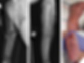
Radiologic technologists have an opportunity to greatly assist the radiologists by performing high quality exams. Unfortunately there is variable quality seen in radiology. (Fig. 1). I explain how these top ten quality tips will greatly reduce errors in the radiology department and improve the ability for radiologists to make an accurate diagnosis. Figure 1. The good, the bad, and the ugly. Unfortunately there is variable quality seen in radiology. Dr. Rice’s Radiography Top Ten: 1. Verify that all images and notes are in PACS for the radiologist. 2. Use a lead side marker with your initials. 3. Remove all clothing, jewelry, and other foreign objects from the field. 4. Use a metal pointer on the area of maximal bone or joint pain. 5. Abdomen x-ray must show both sides of diaphragm and lesser trochanters. 6. Shoulder x-ray must collimate to the shoulder and have 3 views. 7. Wrist X-ray must include a scaphoid view if patient is age 10 or older. 8. CXR must include both lung apices and both lung bases. 9. Accession #, order, and study description for the exam must match the images in PACS. 10. Indication must be available to the radiologist. 1. Verify that all images and notes are in PACS for the radiologist. No sense going to all the trouble to get great images if the radiologist can't see them. If your system does not send an electronic receipt message to the modality, the tech should check the PACS after each case to be sure all the images went through. 2. Use a lead side marker with your initials. The initials are essential so if there is a problem, the radiologist can discuss it with the appropriate person. Technologists should always use physical anatomic side markers placed on the image receptor. Putting on the side marker after taking the image may lead to errors. (Figs. 2-4). This is well documented in the standard textbook for radiographers here: Merrill's Atlas of Radiographic Positioning and Procedures Information on Anatomic Side Markers Figure 2. Electronic side marker, placed on the image with software AFTER it was obtained. The "L" has actually been incorrectly pasted on the patient's right side. Figure 3. Same patient as in Fig. 2 above done the next day, with physical lead side marker placed on the image receptor BEFORE the image was obtained. Technologist has correctly used lead side marker. Figure 4. Example of ambiguous side markers. The radiologist is unable to determine if this is left or right. Study description is LEFT ankle; Marker is RIGHT. If the order is for the wrong side, it is crucial to get the order corrected BEFORE obtaining the radiograph. Also the technologist improperly added the "R" with the imaging software after the image was taken. 3. Remove all clothing, jewelry, and other foreign objects from the field. Extraneous items such as clothing, EKG leads, and jewelry can obscure pathology. (Figs. 5-6) Technologists should be sure to remove it all before taking the image. If the patient refuses or the items can not be removed, the technologist should make a note of it so the radiologist is aware. (Fig. 7. B) Figure 5. Hair extensions on xray. All foreign objects need to be removed prior to taking the radiograph. Figure 6. Jewelry needs to be removed prior to taking the radiograph. Figure 7. A. If there is a reason the images are not following protocol, the tech should make a note of it. Figure 7. B. If the patient refuses or the items can not be removed, the tech should make a note of it. 4. Use a metal pointer on the area of maximal bone or joint pain. The referring physicians have an advantage when they look at the radiographs as they know the exact spot where the patient is having pain. Technologists can give the radiologist the same benefit by placing a metal pointer at the site of pain prior to taking the image. (Figs. 8-10). The technologist should not use the software to insert an electronic pointer after taking the image since this has the same drawbacks as inserting the "L" or "R" after taking the image. (Fig. 10). Using an electronic pointer may result in pointing to the wrong place, or the technologist may be tempted to point to a suspected radiographic abnormality rather than the region of clinical concern. Figure 8. Tech has correctly put a lead marker on the site of maximal tenderness. Figure 9. Same patient as above Fig. 8. Subtle fracture visualized, with help from the tech. Blue arrows are the author's, showing the fracture. Figure 10. A: The tech has put an electronic arrow on the image after it was done. Figure 10. B: The tech has put a physical marker on the site of maximal tenderness. 5. Abdomen x-ray must show both sides of diaphragm and lesser trochanters. Radiologists can miss pathology such as free air or urinary tract calculi if all the anatomy is not included on the images (Fig. 11). Figure 11. A: The upper abdomen is not included on this abdomen radiograph. Figure 11. B: The subsequent chest xray demonstrates free air under the diaphragm (red arrow). Figure 11. C: This is confirmed on the CT scan which shows free air in the abdomen outlining the falciform ligament (blue arrow). 6. Shoulder x-ray must collimate to the shoulder and have 3 views. The central beam should be on the shoulder to avoid geometric distortion. The tech should always shoot a scapular Y view, axillary view, or Grashey view to evaluate for dislocation (Fig. 12). Figure 12. A: The frontal radiograph of the left shoulder demonstrates internal rotation which may give the radiologist a clue to the diagnosis. Figure 12. B: The axillary radiograph of the same patient's left shoulder demonstrates the posterior dislocation and reverse Hill Sachs lesion (also known as a McLaughlin lesion). 7. Wrist X-ray must include a scaphoid view if patient is age 10 or older. Missing a scaphoid fracture may result in non-union and avascular necrosis. Adding the scaphoid view will help the radiologist to make this important diagnosis (Fig. 13). Figure 13: A subtle scaphoid fracture (white arrow) seen on scaphoid view. 8. CXR must include both lung apices and both lung bases. Radiologists can miss pathology such as pneumothorax (Fig. 14), a small pleural effusion, or free air in the abdomen (Fig. 11) if all the anatomy is not included on the images. Figure 14. A: The lower chest is not included on this supine chest radiograph. Figure 14. B: With the lower chest now visible, bilateral pneumothoraces are visualized with a deep sulcus sign on the right (red arrow) and a large lucency on the left (blue arrow). 9. Accession #, order, and study description for the exam must match the images in PACS. All the demographic information has to be correct before sending images to PACS. Trying to fix something after it is in PACS is a nightmare for PACS administrators! 10. Indication must be available to the radiologist. Technologists need to make sure the relevant clinical information is available to the person reading the images. Not only is it important from a patient care perspective, but it is a compliance matter to have a relevant indication on the final report. Through the use of excellent technique, radiographers can greatly assist the radiologists in making an accurate diagnosis, which in turn is a major benefit for our patients. By using the tips in this article you will be on the journey to high quality and safety in the radiology department. Kevin M. Rice, MD serves as the Chair of the Radiology Department and Chief of Staff at Valley Presbyterian Hospital in Los Angeles, California and is a radiologist with Renaissance Imaging Medical Associates. Dr. Rice has made several media appearances as part of his ongoing commitment to public education. Dr. Rice's passion for state of the art radiology and teaching includes acting as a guest lecturer at UCLA. In 2015 Dr. Rice together with Natalie Rice founded Global Radiology CME to provide innovative radiology education at exciting international destinations, with the world's foremost authorities in their field. In 2016, Dr. Rice was nominated and became a semifinalist for a "Minnie" award for the Most Effective Radiology Educator. Follow Dr. Rice on Twitter @KevinRiceMD All posts by Kevin M. Rice, MD
- Large Epiphrenic Esophageal Diverticulum with Achalasia
Progressive dysphagia in a 57 year old male • Xray of the Week This 57-year old male presented with a 4-year history of progressive dysphagia with both solid foods and and liquids. CT scan of the chest with contrast and barium esophagogram were performed. What is the diagnosis? Figure 1. CT scan of the chest with IV contrast. A. Axial image. B. Coronal image. What is the diagnosis? Figure 2. Annotated CT scan of the chest with IV contrast. A. Axial image demonstrates a large outpouching (red arrow) with an air fluid level, due to the large esophageal diverticulum. B. Coronal image clearly shows the large right-sided esophageal diverticulum (red arrow) which communicates with the esophagus (green arrow). The esophagus is also dilated (yellow arrow). Figure 3. Esophagram depicts the presence of a large right-sided diverticulum (red arrow) approximately 8 cm from the gastroesophageal junction. There is pooling of the contrast inside the diverticulum as it communicates with the esophagus. The esophagus is also dilated with a characteristic “bird’s beak appearance”/tapering (yellow arrow) near the gastroesophageal junction. Discussion: Epiphrenic esophageal diverticulum (EED) is an uncommon form of esophageal diverticulum with an incidence of only 1:500,000/year in the adult population, and comprises about 10% of all esophageal diverticula. Arising in the distal esophagus, it is typically located 4 to 8 cm above the cardia or in the last 10 cm of the esophagus (1-3). It is a pulsion type of diverticulum caused by motility disorders with markedly increased intraluminal esophageal pressures (1-2). Presentation in infants and children is rarely seen (4). In addition to other motility disorders, achalasia is almost always associated with EED (5). Achalasia itself is a rare entity with an approximate incidence of 1:100,000 people/year (6-7). Clinical manifestations of EED are variable but most commonly include dysphagia and regurgitation which usually progress over time (8). However, up to eighty percent of patients may be asymptomatic. Diagnosis is usually made via barium swallow with supplemental evaluation including upper endoscopy to exclude malignancy and esophageal manometry to delineate any underlying motility disorder (9). Treatment strategies, either open or laparoscopic surgery, involve esophageal myotomy, diverticulectomy and fundoplication. Myotomy decreases the incidence of diverticulum recurrence and partial fundoplication has been proposed as the most appropriate anti-reflux method. Surgery appears to be an effective treatment but is associated with significant mortality and morbidity, thus is it reserved for patients with symptomatic EED (10). References: 1. Debas HT, Payne WS, Cameron AJ, et al (1980). Physiopathology of lower esophageal diverticulum and its implications for treatment. Surg Gynecol Obstet; 151:593–600. 2. Dodds WJ, Stef JJ, Hogan WJ, et al (1975). Radial distribution of peristaltic pressure in normal subjects and patients with esophageal diverticulum. Gastroenterology; 69:584–590. 3. Clouse RE, Diamant NE (2006). From Esophageal motor and sensory function and motor disorders of the esophagus. Sleisenger and Fordtran’s Gastrointestinal and Liver disease. Edited by Feldman M, Freeman LS, Brandt LJ. Philadelphia, WB Saunders; 855-904. 4. Zaninotto G, Portale G, Costantini M, et al (2011). Therapeutic strategies for epiphrenic diverticula: Systematic review. World J Surg; 35:1447-53. 5. Fisichella PM, Jalilvand A, Dobrowolsky A (2015). Achalasia and epiphrenic diverticulum. World J Surg.; 39 (7):1614-9. 6. Vaezi MF, Richter JE (1999). Diagnosis and management of achalasia. American College of Gastroenterology Practice Parameter Committee. Am J Gastroenterol; 94: 3406 – 12. 7. Francis DL, Katzka DA (2010). Achalasia: update on the disease and its treatment. Gastroenterology; 139: 369 – 74. 8. Thomas ML, Anthony A, Fosh BG, et al (2001). Esophageal diverticula. Br J Surg; 88: 629-42. 9. Fasano NC, Levine MS, Rubesin SE, et aI (2003). Epiphrenic diverticulum: clinical and radiographic findings in 27 patients. Dysphagia; 18 (1):9e15. 10. Benacci JC, Deschamps C, Trastek VF, et al (1993). Epiphrenic diverticulum: results of surgical treatment. Ann Thorac Surg; 55:1109 –1114. Karl John A. Koa, MD completed his Doctor of Medicine at West Visayas State University, in La Paz, Iloilo City, Philippines. He is currently the senior and Chief Resident Physician in the Department of Radiology at Chong Hua Hospital, also in Philippines. He has presented case reports in the West Visayas State University including a case report contest on Neurofibromatosis Type 2, which he won last 2015 during his post graduate internship training.
- Prof. MUDr. Jiří Ferda MD, Ph.D.- President of the Czech Radiological Society Joins Global Radiology
Global Radiology CME is pleased to welcome Prof. MUDr. Jiri Ferda, President of the Czech Radiological Society, Deputy-Head of Department of Imaging, Head of Molecular Imaging Center, Professor of Radiology - Medical Faculty Pilsen, Charles University, Czech Republic to Global Radiology's world renowned faculty for Imaging in Prague. Professional Qualification Diploma in Radiology, Diploma in Neuroradiology, Diploma in Interventional Radiology and Diploma in Nuclear Medicine Professional orientation: Vascular diagnostic and intervention radiology, Computed tomography - advanced CT applications, Magnetic resonance imaging – neuroimaging, cardiac, vascular and abdominal imaging, molecular and hybrid imaging PET/CT, PET/MRI Publications and presented lectures More than 100 scientific publications, 5 monographies, 7 chapters in monography Selected publications: Ferda J, Novák M, Mírka H, Baxa J, Ferdová E, Bednárová A, Flohr T, Schmidt B, Klotz E, Kreuzberg B. The assessment of intracranial bleeding with virtual unenhanced imaging by means of dual-energy CT angiography. Eur Radiol. 2009 Oct;19(10):2518-22. Ferda J, Ferdová E, Záhlava J, Matejovic M, Kreuzberg B. Fever of unknown origin: a value of (18)F-FDG-PET/CT with integrated full diagnostic isotropic CT imaging. Eur J Radiol. 2010 Mar;73(3):518-25. Ferda J, Ferdová E, Mírka H, Baxa J, Bednářová A, Flohr T, Schmidt B, Matějovič M, Kreuzberg B. Pulmonary imaging using dual-energy CT, a role of the assessment of iodine and air distribution. Eur J Radiol. 2011 Feb;77(2):287-93 Ferda J, Ferdová E, Hes O, Mraček J, Kreuzberg B, Baxa J. PET/MRI: Multiparametric imaging of brain tumors. Eur J Radiol. 2017 Sep;94:A14-A25. Ferda J, Hromádka M, Baxa J. Imaging of the myocardium using (18)F-FDG-PET/MRI. Eur J Radiol. 2016 Oct;85(10):1900-1908.
- Congenital Syphilis
6 Week Old Baby with Skin Rash and Bilateral Bone Abnormalities • Xray of the Week What is the diagnosis? Figure 1. Radiographs of the upper and lower extremities. Clinical images of the hand and foot. What is the diagnosis? Clinical images courtesy of Peter Koetters, MD. Figure 2. Annotated radiographs of the upper and lower extremities. There is diffuse mild periostitis of the long bones (red arrows). Mild irregularity of the distal femoral and proximal tibial metaphyses is also present (green arrows). There is mild sclerosis and cupping of the distal radial and ulnar metaphyses as well as the proximal fibular metaphysis (yellow arrows). A rash on the hands and soles with associated with desquamation is another clue to the diagnosis. Clinical images courtesy of Peter Koetters, MD. Discussion: Congenital syphilis is still a major problem in sub-Saharan Africa, but there is a resurgence in several European countries and in North America. Congenital syphilis occurs when Treponema pallidum crosses the placenta during birth or by contact with an infectious lesion. Mucocutaneous involvement is present in about 70% of infants with early congenital syphilis. It is typically a vesicular or maculopapular rash occurring on the palms and soles. As seen in this case, it may be associated with desquamation. The musculoskeletal anomalies include periostitis, metaphysitis, sawtooth metaphysis, diaphyseal osteomyelitis, pathological fractures, joint effusions, sabre shin, dactylitis, and several craniofacial anomalies. Treatment is with benzathine penicillin G, and prognosis is usually good unless non-reversible changes have occured. References: 1. Rasool MN, Govender S. The Skeletal Manifestations of Congenital Syphilis: A Review Of 197 Cases from the University of Natal. J Bone Joint Surg [Br] 1989 :7 I-B :752-755. https://online.boneandjoint.org.uk/doi/pdf/10.1302/0301-620x.71b5.2584243 2. Ferreira ST, Correia C, Marçal M, et al. Skin rash: a manifestation of early congenital syphilis. BMJ Case Rep Published online: 12 May 2016 doi:10.1136/ bcr-2016-216148 3. Hook EW, Peeling RW. Syphilis control--a continuing challenge. The New England Journal of Medicine. 2001 July. 351(2):122-124. doi:10.1056/NEJMp048126 4. Russo PE, Shryock LF. Bone lesions of congenital syphilis in infants and adolescents: report of 46 cases. Radiology. 44(5):477-84. 5. Phiske MM. Current trends in congenital syphilis. Indian J Sex Transm Dis AIDS. 2014 Jan-Jun; 35(1): 12–20. doi: 10.4103/2589-0557.132404 6. Gupta R, Vora R. Congenital syphilis, still a reality. Indian J Sex Transm Dis AIDS. 2013 Jan-Jun; 34(1): 50–52.doi: 10.4103/2589-0557.112941 Kellie Greenblatt, MD Pediatric Radiologist, RIMA A native of the Bay Area, Dr. Greenblatt attended the University of California at Berkeley where she earned a BA in Human Biodynamics. Following graduation, Dr. Greenblatt performed basic research for four years in the toxicology laboratory at Lawrence Livermore Laboratory focusing on carcinogens found naturally in cooked meat. Following her work at Lawrence Livermore, Dr. Greenblatt attended medical school at the Chicago Medical School. After medical school, Dr. Greenblatt did two years of residency training in Surgery before entering a Diagnostic Radiology residency. She completed her Radiology Residency at Kaiser Permanente in Los Angeles, California, then did a one year fellowship in Pediatric Radiology at Children’s Hospital Los Angeles. Kevin M. Rice, MD is president of Global Radiology CME and serves as the Chief of staff and Chair of the Radiology Department of Valley Presbyterian Hospital in Los Angeles, California and is a radiologist with Renaissance Imaging Medical Associates. Dr. Rice's passion for state of the art radiology and teaching includes acting as a guest lecturer at UCLA. Dr. Rice co-founded Global Radiology CME with Natalie Rice to provide innovative radiology education at exciting international destinations, with the world's foremost authorities in their field. In 2016, Dr. Rice was nominated and became a semifinalist for a "Minnie" Award for the Most Effective Radiology Educator. Follow Dr. Rice on Twitter @KevinRiceMD All posts by Kevin M. Rice, MD
- Josef Vymazal - Prominent Czech Radiologist Joins Global Radiology's World Renowned Faculty for
Representatives from Global Radiology CME visited Prague the week of September 24, 2018 to work on the final preparations for Imaging in Prague 2019. We had the pleasure of meeting Dr. Josef Vymazal, Head, Department of Radiology at Na Homolce Hospital, Full Professor at Charles University, Prague, Czech Republic to our faculty. Dr. Vymazal is a neuroradiologist who trained in the USA at the NIH. He is also a practicing neurologist and the director of Na Homolce Hospital in Prague, a neurology and cardiac specialty center. In 2014-2016, Dr. Vymazal served as Deputy Minister of Health for the Czech Republic, and is currently an Adviser to Minister of Health in the Czech Republic. Dr. Vymazal will join our prestigious team that includes, Dr. Donald Resnick, MSK expert, and 2018 recipient of the ACR Gold Medal, Dr. Phillip Tirman, MSK expert, a 2018 Aunt Minnie semifinalist, in the category of most Effective Radiology Educator, Dr. Blake Johnson, highly sought Neuroradiologist lecturer, and Medical Director and Director of Neuroimaging at Center for Diagnostic Imaging, Dr. Neil Rofsky, world renowned body imager, past president SCBTMR, and currently, Professor and Chair of Radiology and the Effie and Wofford Cain Distinguished Chair in Diagnostic Imaging of University of Texas Southwestern. We are also honored to be adding Dr. Paul Parizel, Past President of the ESR and Chairman of the Department of Radiology at the Antwerp University Hospital, and tenured Professor of Radiology, University of Antwerp, Belgium as well as Dr. Nicole Hindman, Associate Professor - Departments of Radiology and Surgery and Director, Female Pelvic Imaging at New York University. We welcome you to join Radiologists from around the world to experience academic excellence and networking and social opportunities the city renowned as the Architectural Jewel of World, rich in history, with a vibrant energy! Natalie Rice, Kevin Rice, and Josef Vymazal in Prague