


Papillary Fibroelastoma of Aortic Valve
- Kevin M. Rice, MD

- Oct 10, 2015
- 2 min read
Updated: Jul 26, 2021
57 yo Female with Intermittent Chest Pain • Xray of the Week
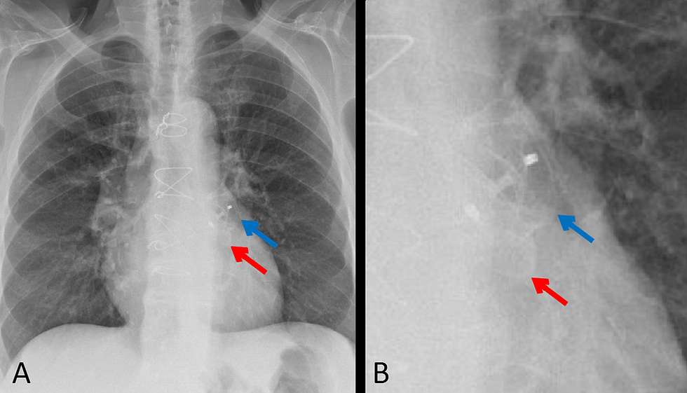
This 57 year old female presented with atypical chest pain and an echocardiogram showed an abnormality of the aortic valve. Blood cultures and cardiac enzymes were both normal.

Coronal CT (left panel) and axial (right panel) images showing lobulated mass arising from the right cusp of the aortic valve.
Papillary fibroelastomas are the third most common type of primary tumor of the heart, behind cardiac myxomas and cardiac lipomas, and are the most common benign neoplasms of the cardiac valvular structures. Papillary fibroelastomas may cause chest pain due to intermittent occlusion of the coronary arteries. Fragments of the tumor may embolize and cause stroke if they enter the intracranial circulation. Myocardial infarction or sudden cardiac death may be due to embolization of a portion of the tumor into a coronary artery. Surgical resection should be considered for all patients who have symptoms and for asymptomatic patients who have pedunculated lesions or tumors larger than 1 cm. Valve-sparing excision usually results in good long-term results.
References:
1. Sun, JP, et al. Clinical and Echocardiographic Characteristics of Papillary Fibroelastomas: A Retrospective and Prospective Study in 162 Patients. Circulation.2001; 103: 2687-2693
2. Kumbala D, Sharp T, Kamalesh M. "Perilous pearl"-papillary fibroelastoma of aortic valve: a case report and literature review. Angiology. 2008;59 (5): 625-8.
3. Araoz PA, Eklund HE, Welch TJ et-al. CT and MR imaging of primary cardiac malignancies. Radiographics. 1999;19 (6): 1421-34.
4. Lembcke A, Meyer R, Kivelitz D, et al. Papillary Fibroelastoma of the Aortic Valve. Appearance in 64-Slice Spiral Computed Tomography, Magnetic Resonance Imaging, and Echocardiography. Circulation. 2007;115:e3-e6
5. Gopaldas RR, Atluri PV, Blaustein AS, Bakaeen FG, Huh J, Chu D. Papillary fibroelastoma of the aortic valve: operative approaches upon incidental discovery. Tex Heart Inst J. 2009; 36(2): 160–163.

Kevin M. Rice, MD is the president of Global Radiology CME
Dr. Rice serves as the Chair of the Radiology Department of Valley Presbyterian Hospital in Los Angeles, California and is a radiologist with Renaissance Imaging Medical Associates. Dr. Rice has made several media appearances as part of his ongoing commitment to public education. Dr. Rice's passion for state of the art radiology and teaching includes acting as a guest lecturer at UCLA. In 2015 Dr. Rice launched Global Radiology CME to provide innovative radiology education at exciting international destinations, with the world's foremost authorities in their field.
Follow Dr. Rice on Twitter at @KevinRiceMD


Comments