


Amulet® Left Atrial Appendage Closure Device
- Kevin M. Rice, MD

- Feb 17, 2020
- 3 min read
Updated: Jul 26, 2021
Name the Cardiac Device • Xray of the Week
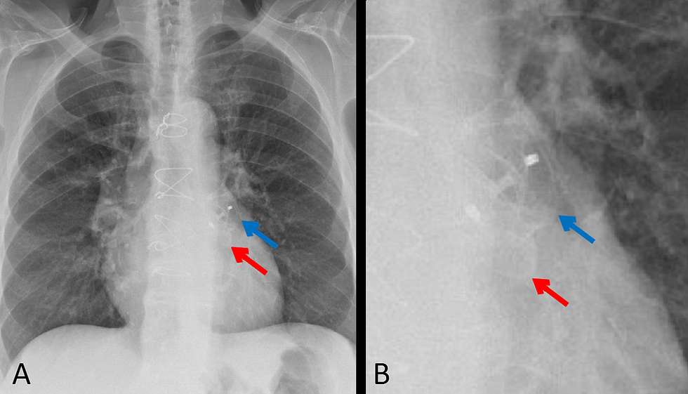
Fig. 1. A. Frontal chest xray showing the Amulet® over the expected location of the left atrial appendage (LAA). Fig. 1. B. Magnified chest xray. The self-expanding distal lobe (blue arrow) and proximal disc (red arrow) are seen.
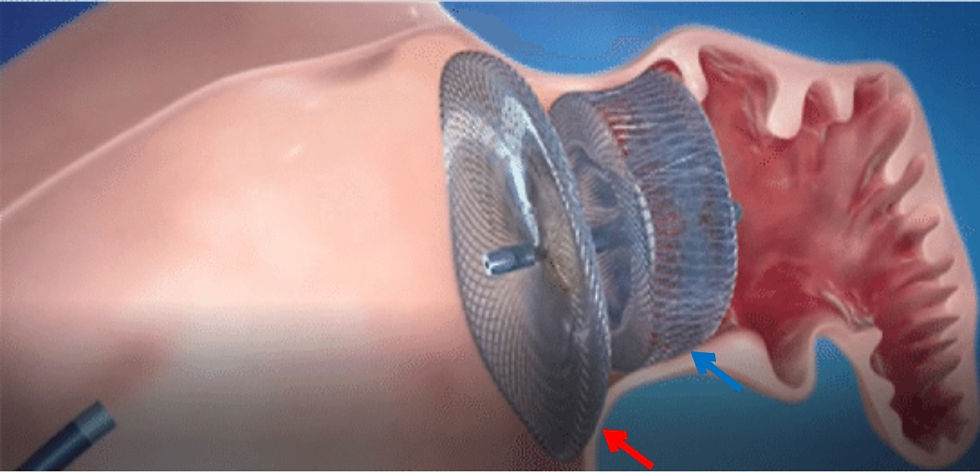
Fig. 2. Amulet® is very similar to the Amplatzer™ Cardiac Plug (ACP) device.
Fig. 3 Video demonstrating percutaneous placement technique for the Amplatzer™ Cardiac Plug (ACP) and Amulet® device for LAA closure.
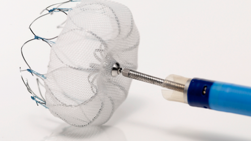
Fig. 4. Watchman™ LAAC device from Boston Scientific shows the self-expanding nitinol frame and fabric covering the face of the device.

Fig. 5. CT scan of Watchman™ LAAC device in the left atrial appendage on axial and coronal images.
Fig. 6 Video explaining percutaneous placement technique for the Watchman™ LAAC device.

Fig. 7. AtriClip® Left Atrial Appendage Exclusion System. A. The AtriClip® in the deployment device. B. The layers of the AtriClip®.
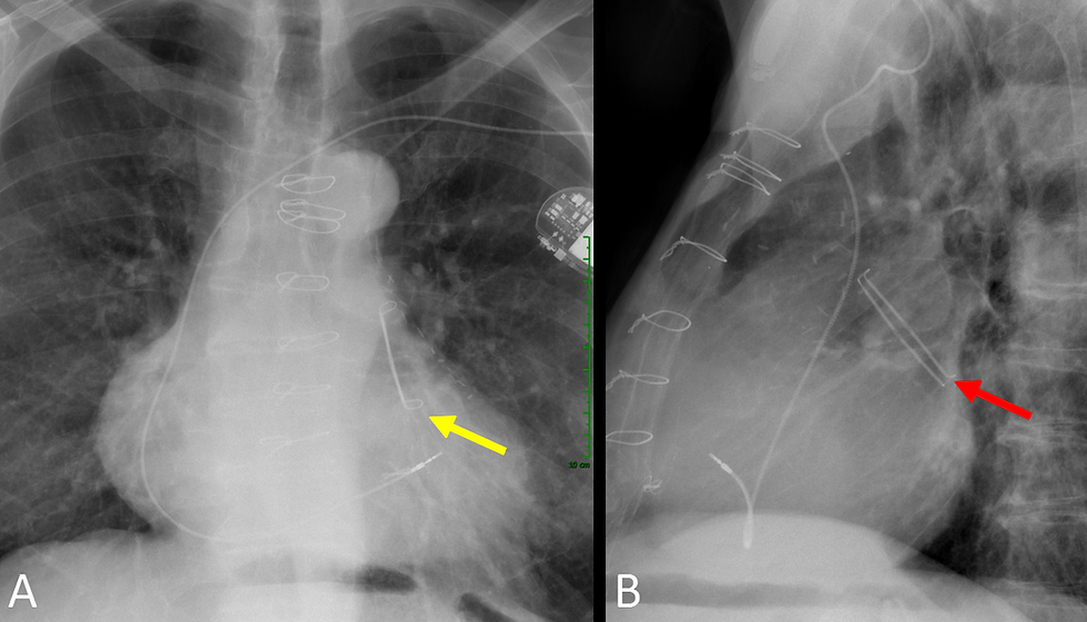
Fig. 8. AtriClip® Left Atrial Appendage Exclusion System. A. Frontal CXR with yellow arrow pointing to the device. B. Lateral CXR with red arrow pointing to the AtriClip®. Note the parallel tubes and Nitinol springs at each end.
Discussion:
LAA closure or occlusion devices are indicated for patients with atrial fibrillation in whom oral anticoagulation is contraindicated or an alternative to oral anticoagulation therapy for stroke prevention in patients with atrial fibrillation. Percutaneous LAA closure devices include the Watchman, Amplatzer Amulet, Amplatzer Cardiac Plug (ACP), and the PLAATO system. The Amulet and ACP both consist of a self-expanding distal lobe and proximal disc made of nitinol mesh with an articulating waist (Figs 1,2,3). The Watchman consists of a self-expanding nitinol frame, with a fabric covering the face of the device (Figs 4-6). Placed by open surgery or minimally invasive techniques, the AtriClip (Fig 7,8) is a self-closing clip placed on the epicardial surface of the heart on the base of the LAA . It is visualized on radiographs as a metallic structure with parallel tubes over the expected location of the LAA (Fig 6,7).
Complications of percutaneous LAA closure devices include malposition, migration, or embolization. The Watchman, AtriClip, Amplatzer Amulet, and Amplatzer Cardiac Plug LAA closure devices are MR imaging conditional at 1.5 T and 3 T.
References:
1. Swaans MJ, Wintgens LI, Alipour A, Rensing BJ, Boersma LV. Percutaneous left atrial appendage closure devices: safety, efficacy, and clinical utility. Med Devices (Auckl). 2016;9:309-316. Published 2016 Sep 2. doi:10.2147/MDER.S65492 Full Text: https://www.ncbi.nlm.nih.gov/pmc/articles/PMC5015878/
2. Onalan O, Crystal E. Left atrial appendage exclusion for stroke prevention in patients with nonrheumatic atrial fibrillation. Stroke. 38 (2 Suppl): 624-30. Full Text: doi:10.1161/01.STR.0000250166.06949.95
3. Sigakis CJG, Mathai SK, Suby-Long TD, Restauri NL, Ocazionez D, Bang TJ, Restrepo CS, Sachs PB, Vargas D. Radiographic Review of Current Therapeutic and Monitoring Devices in the Chest. (2018) Radiographics : a review publication of the Radiological Society of North America, Inc. 38 (4): 1027-1045. doi:10.1148/rg.2018170096 Full Text: https://pubs.rsna.org/doi/10.1148/rg.2018170096
4. Bedeir K, Warriner S, Kofsky E, Gullett C, Ramlawi B. Left Atrial Appendage Epicardial Clip (AtriClip): Essentials and Post-Procedure Management. (2019) Journal of atrial fibrillation. 11 (6): 2087. doi:10.4022/jafib.2087 Full Text: https://www.ncbi.nlm.nih.gov/pmc/articles/PMC6652788/
5. Moussa Pacha H, Al-Khadra Y, Soud M, Darmoch F, Moussa Pacha A, Alraies MC. Percutaneous devices for left atrial appendage occlusion: A contemporary review. World J Cardiol. 2019;11(2):57–70. doi:10.4330/wjc.v11.i2.57 Full Text: https://www.ncbi.nlm.nih.gov/pmc/articles/PMC6391622/
6. Sick PB, Schuler G, Hauptmann KE, et al. Initial worldwide experience with the WATCHMAN left atrial appendage system for stroke prevention in atrial fibrillation. J Am Coll Cardiol. 2007;49(13):1490-1495. doi:10.1016/j.jacc.2007.02.035. Full Text: https://www.sciencedirect.com/science/article/pii/S0735109707007474
Related posts:

Kevin M. Rice, MD is the president of Global Radiology CME
Dr. Rice is a radiologist with Renaissance Imaging Medical Associates. Dr. Rice has made several media appearances as part of his ongoing commitment to public education. Dr. Rice's passion for state of the art radiology and teaching includes acting as a guest lecturer at UCLA. In 2015, Dr. Rice and Natalie Rice founded Global Radiology CME to provide innovative radiology education at exciting international destinations, with the world's foremost authorities in their field.
Follow Dr. Rice on Twitter @KevinRiceMD


Comments