


Amplatzer® Septal Occluder in ASD
- Neal Shah and Kevin M. Rice, MD
- Apr 12, 2020
- 4 min read
Updated: Jan 7, 2023
Name the Cardiac Device • Xray of the Week

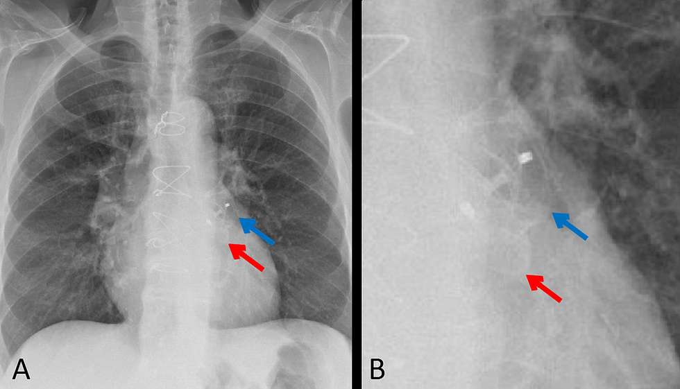
Fig. 1. A. Frontal chest xray showing the Amplatzer® Septal Occluder Device over the expected location of the atrial septum (red arrows).
Fig. 1. B. Echochardiogram 4 chamber view showing the Amplatzer® Septal Occluder Device (yellow arrows) covering both sides of the atrial septum.
Fig. 1. C. CT scan showing the Amplatzer® Septal Occluder Device (green arrows) covering both sides of the atrial septum. The self-expanding distal lobe is seen in the left atrium and the proximal disc is seen in the right atrium. A large pericardial effusion is also present.

Fig. 2. Frontal (A) and lateral (B) chest xray in a different patient showing the Amplatzer® Septal Occluder Device over the expected location of the atrial septum (red arrows).

Fig. 3. Amplatzer® Septal Occluder device.
Fig. 4 Video demonstrating percutaneous placement technique for the Amplatzer® Septal Occluder for ASD closure.
Discussion:
The Amplatzer Septal Occluder (ASO) is designed for percutaneous closure of atrial septal defect (ASD), the fourth most common congenital cardiac anomaly. ASD closure is indicated within the setting of right cardiac chamber enlargement, prevention of paradoxical embolism, net left to right shunting, and to prevent arrythmias [1]. The device is shaped as a self-expanding double disc composed of a nitinol mesh with polyester fabric (Figs.1-3). The double disc shape allows closure from both sides of the septal defect with one disc being placed alongside the left septal wall within the left atrium, and the other being placed along the right septal wall within the right atrium. This means that only ASD secundum type defects are able to be repaired with this device [2,3]. Despite the improving accuracy of both 2D and 3D echocardiography, fluoroscopy remains the standard for periprocedural imaging [4,5]. After the device has been placed postprocedural imaging is typically conducted via echocardiography to evaluate for device positioning and any residual shunting. As seen in this case, the discs are visible over the interatrial septum on radiographs, ultrasound, and CT scan (Figs.1,2). Closure with this device is a highly successful procedure and offers lower rates of post-procedural complications than seen with open heart surgery and a shorter hospital stay (Fig. 4). The most common complication during placement is device embolization or malposition which occurs in 3.5% of cases. The most frequent long term complication following ASO placement is arrythmias, typically supraventricular tachyarrhythmias [6]. The other potential long term complication is myocardial erosion which may lead to pericardial effusion or tamponade [6]. Mortality rates between the ASO device and surgical groups in studies tend to be similar [6].
References:
Holland M. Amplatzer septal occluder | Radiology Case | Radiopaedia.org. Radiopaedia. https://radiopaedia.org/cases/amplatzer-septal-occluder?lang=us. Accessed April 12, 2020.
Kim H-H, Yi G-J, Song S-W. Late Migration of Amplatzer Septal Occluder Device to the Descending Thoracic Aorta. Korean J Thorac Cardiovasc Surg. 2017;50(1):47-49. doi:10.5090/kjtcs.2017.50.1.47
Sigakis CJG, Mathai SK, Suby-Long TD, et al. Radiographic Review of Current Therapeutic and Monitoring Devices in the Chest. RadioGraphics. 2018;38(4):1027-1045. doi:10.1148/rg.2018170096
Sigakis CJG, Mathai SK, Suby-Long TD, et al. Radiographic Review of Current Therapeutic and Monitoring Devices in the Chest. RadioGraphics. 2018;38(4):1027-1045. doi:10.1148/rg.2018170096
Ackermann S, Quandt D, Hagenbuch N, et al. Transcatheter Atrial Septal Defect Closure in Children with and without Fluoroscopy: A Comparison. Journal of Interventional Cardiology. doi:https://doi.org/10.1155/2019/6598637
Spence MS, Qureshi SA. Complications of transcatheter closure of atrial septal defects. Heart. 2005;91(12):1512-1514. doi:10.1136/hrt.2004.057562
Related posts:

Neal Shah went to medical school at The Edward Via College of Osteopathic Medicine (VCOM)–Carolinas and matched in radiology at Vanderbilt University Medical Center. Prior to medical school, he completed his undergraduate studies at the University of North Carolina at Chapel Hill where he majored in economics and chemistry. During his 4 years there he worked in UNC’s Biomedical Research Imaging Center where he helped develop formulations for iron-oxide nanoparticles used for MRI; it was here that his love for the field of radiology developed. He eventually wishes to also pursue his MBA and hopes to use it to help advance the field of medicine in terms of medical innovation.
Follow Neal Shah on Twitter @neal-shah17

Kevin M. Rice, MD is the president of Global Radiology CME
Dr. Rice is a radiologist with Cape Radiology Group, and formerly the Chief of Staff at at Valley Presbyterian Hospital in Los Angeles, California. Dr. Rice has made several media appearances as part of his ongoing commitment to public education. Dr. Rice's passion for state of the art radiology and teaching includes acting as a guest lecturer at UCLA.In 2015, Dr. Rice and Natalie Rice founded Global Radiology CME to provide innovative radiology education at exciting international destinations, with the world's foremost authorities in their field. In 2016, Dr. Rice was nominated and became a semifinalist for a "Minnie" Award for the Most Effective Radiology Educator.He was once again a semifinalist for a "Minnie" for 2021's Most Effective Radiology Educator by AuntMinnie.com.
Follow Dr. Rice on Twitter @KevinRiceMD


Comments