Traumatic Thoracic Aortic Injury
- Han Ngo and Kevin M. Rice, MD
- Jun 30, 2020
- 4 min read
Updated: May 7, 2021
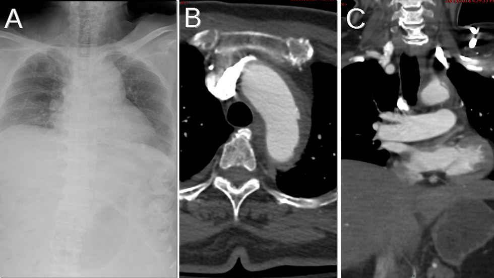
48 year old male with chest pain following a motor vehicle collision • Xray of the Week

Figure 1. Name the important findings on this CXR and CT Scan.

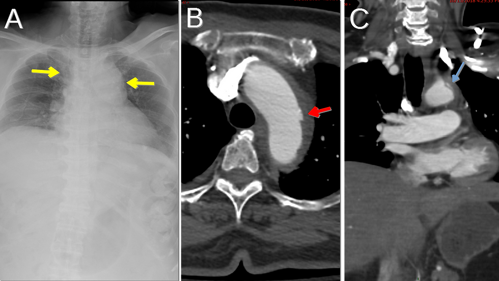
Figure 2. Imaging results of a trauma patient who was in a motor vehicle collision. A: Chest X-ray demonstrating a widening of the mediastinum (yellow arrows). B: Axial CT image of the chest showing subtle pseudoaneurysm along the lateral aspect of the aortic arch (red arrow). C: Coronal CT image of the chest showing hematoma adjacent to the site aortic injury (blue arrow).

Figure 3. Same patient as in figure 2, one month after aortic stent graft. A: Chest X-ray showing a normal width of the mediastinum after the placement of the stent graft (yellow arrow). B & C: Axial and coronal CT images of the chest showing a good expansion of the stent without adjacent hematoma or leak (red and blue arrows).

Figure 4. Traumatic Aortic Injury Grading System (5)- Diagram by Han Ngo
Grade 1: Intimal flap/intimal tear.
Grade 2: Intramural hematoma (hemorrhage without an intimal tear).
Grade 3: Aortic pseudoaneurysm (false aortic rupture contained by the thin wall of adventitia).
Grade 4: Aortic transection (true aortic rupture due to the damage of all three layers).
Discussion:
Thoracic aortic injury is the most common type of traumatic aortic injury (TAI), a life-threatening condition that frequently occurs as a result of crush or deceleration injuries. As the second most common cause of death in patients with blunt trauma, TAI has a very high mortality rate with up to 80% of patients die at the scene of trauma and of those who survive the arrival to the ER, 30% die within the first 24 hours (1). Early diagnosis of TAI relies on appropriate imaging since clinical symptoms and examination are nonspecific. Chest X-ray can detect indirect signs of TAI such as widened mediastinum (Figs. 1,2), trachea displacement, depression of left main bronchus, obliteration of aortic knob contour, left apical pleural cap, or hemothorax. Although nonspecific, these findings should raise high suspicion for TAI when present in blunt trauma cases. CT scan can reveal direct signs of TAI through the visualization of aortic injuries (Figs. 1,2). Aortic injuries are categorized into four grades based on severity: intimal flap, intramural hematoma, pseudoaneurysm, or aortic transection (2-5) (Fig. 4). This is a case of Grade 3 injury due to the presence of a pseudoaneurysm (Fig. 1,2).
Aortic stent graft (Fig. 3) is the preferred treatment for TAI because of its higher success rate and lower rate of complication than open surgical repair. The stent is placed by inserting a catheter with the compressed stent through an artery, most commonly the femoral artery. Once the stent reaches the site of aortic lesion, it is expanded to form a new aortic wall and prevent more blood from entering the pseudoaneurysm (3-5).
References:
1. Igiebor OS, Waseem M. Aortic Trauma. StatPearls Publishing LLC. https://www.ncbi.nlm.nih.gov/books/NBK459337/. Published December 16, 2019. Accessed June 30, 2020.
2. Yahia DAA, Bouvier A, Nedelcu C, et al. Imaging of thoracic aortic injury. Diagn Interv Imaging. 2015;96(1):79-88. doi:10.1016/j.diii.2014.02.003
3. Mokrane FZ, Revel-Mouroz P, Saint Lebes B, Rousseau H. Traumatic injuries of the thoracic aorta: The role of imaging in diagnosis and treatment. Diagn Interv Imaging. 2015;96(7-8):693-706. doi:10.1016/j.diii.2015.06.005
4. Yamane BH, Tefera G, Hoch JR, et al. Blunt thoracic aortic injury: Open or stent graft repair? Surgery. 2008;144(4):575-582. doi:10.1016/j.surg.2008.06.007
5. Azizzadeh A, Keyhani K, Miller CC, et al. Blunt traumatic aortic injury: Initial experience with endovascular repair. J Vasc Surg. 2009 Jun;49(6):1403-8. doi:10.1016/j.jvs.2009.02.234

Han Ngo is a medical student at Oakland University William Beaumont School of Medicine (OUWB) in Rochester, Michigan. She graduated from UCLA, receiving her B.S. degree in Biochemistry. Prior to starting medical school, Han spent 4+ years (including her undergraduate years) working as a medical scribe for a psychiatrist at Ronald Reagan UCLA Medical Center. Interested in radiology, Han is now serving as the President of both diagnostic radiology and interventional radiology interest groups at OUWB. She is also a committee member on the Medical Student Council of the Society of Interventional Radiology (SIR). After deciding on her specialty, Han plans to continue learning and striving to make a difference in patients’ lives.
Follow Han Ngo on Twitter @Han_Ngoo

Kevin M. Rice, MD is the president of Global Radiology CME
Dr. Rice is a radiologist with Renaissance Imaging Medical Associates and is currently the Vice Chief of Staff at Valley Presbyterian Hospital in Los Angeles, California. Dr. Rice has made several media appearances as part of his ongoing commitment to public education. Dr. Rice's passion for state of the art radiology and teaching includes acting as a guest lecturer at UCLA. In 2015, Dr. Rice and Natalie Rice founded Global Radiology CME to provide innovative radiology education at exciting international destinations, with the world's foremost authorities in their field. In 2016, Dr. Rice was nominated and became a semifinalist for a "Minnie" Award for the Most Effective Radiology Educator.
Follow Dr. Rice on Twitter @KevinRiceMD


Comments