


Type 1 Dens Fracture
Trauma due to motor vehicle collision. Diagnosis? • Xray of the Week

Figure 1. CT images of the cervical spine (A) sagittal view and (B) coronal view. What is the important finding on this CT scan?
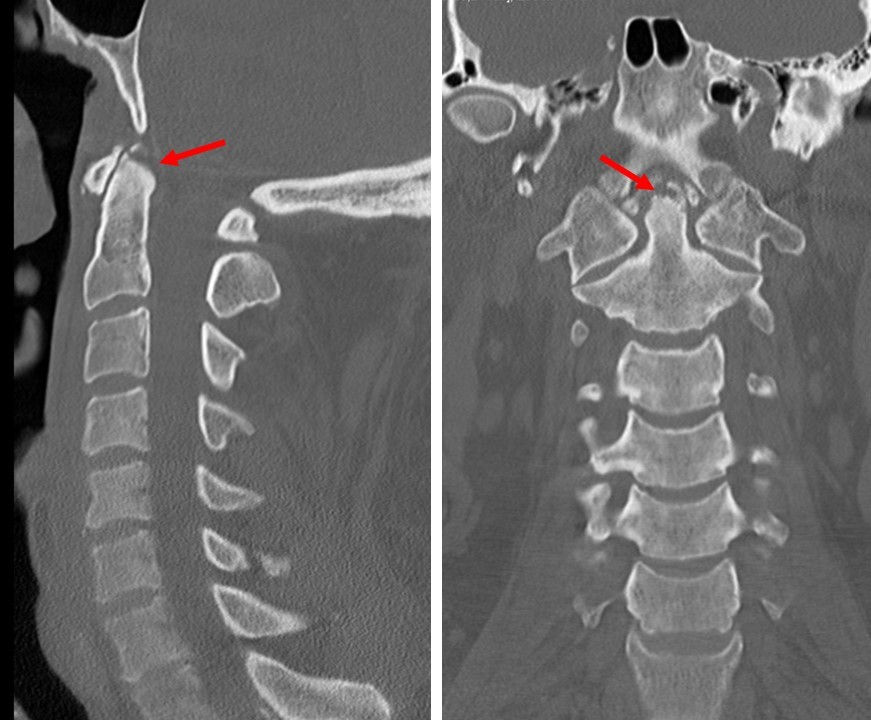
Figure 2. CT scan of a Type I dens fracture. Red arrows point to the fracture line at the tip of the dens.

Figure 3. Anderson and D’Alonzo dens fracture classification system. Diagram by Neal Joshi.
Type I is an oblique avulsion fracture involving the upper tip of the dens, usually at the alar ligament insertion, and is generally considered stable when isolated.
Type II occurs at the junction of the dens and the body of C2 and has a greater risk of displacement and nonunion.
Type III extends from the base of the dens into the body of C2 and generally has better healing potential because the fracture extends into cancellous bone.
Introduction:
The odontoid process, or dens, is a superior projection of C2 that articulates with the anterior arch of C1 and functions as the pivot for atlantoaxial rotation. The alar ligaments arise from the upper dens and attach to the medial aspects of the occipital condyles, contributing to stability of the craniocervical junction (1,2). The Anderson and D'Alonzo classification is the most widely used system for classifying odontoid fractures according to the location of the fracture (3,4) (Fig. 3). Type I fracture is usually stable, however, an apparent Type I avulsion fracture may be associated with injury to the alar or other craniocervical ligaments; therefore, the surrounding ligamentous structures and craniocervical alignment should be assessed (5).
Epidemiology
Odontoid fractures are among the most common fractures of the upper cervical spine, particularly in older adults. Type I odontoid fractures are rare, representing approximately 2–3% of odontoid fractures. In a prospective study from the Swedish Fracture Register, 30 of 1,154 odontoid fractures were classified as Type I, compared with 583 Type II and 541 Type III fractures. Odontoid fractures in older adults are commonly associated with low-energy falls, whereas higher-energy mechanisms such as motor vehicle collisions are more frequent in younger patients (6,7).
Clinical Manifestation
Patients with Type I dens fractures typically present with upper cervical or posterior neck pain and tenderness following trauma. Pain may be aggravated by cervical movement and may be associated with restricted range of motion. Neurological deficits are uncommon but may occur in the presence of associated cervical or craniocervical injuries. Because Type I fractures are uncommon and may be associated with ligamentous injury, careful clinical and radiologic assessment for craniocervical instability is important (3).
Radiographic Features
Plain radiographs, including lateral and open-mouth odontoid views, may demonstrate a small avulsion fragment at the tip of the dens. However, these fractures may be subtle and difficult to detect on conventional radiographs. For odontoid fractures, radiographs can be very useful, but a negative result does not exclude a fracture. Therefore, if there is clinical suspicion a CT scan should be obtained (Fig. 1). Computed tomography (CT) is the preferred imaging modality for evaluating suspected odontoid fractures. Thin-section CT with sagittal and coronal reformations allows accurate assessment of the fracture location, displacement, and alignment of the atlantoaxial and craniocervical junctions. CT scan of this patient following a motor vehicle collision shows fracture involving the tip of the dens (Fig. 2). Associated fractures of C1 and C2 should also be carefully evaluated (5). MRI may be useful when there is concern for associated ligamentous injury or craniocervical instability and can provide additional evaluation of the alar, apical, and transverse ligaments as well as the spinal cord.
Treatment and Prognosis
Isolated, nondisplaced Type I dens fractures are generally considered stable and are treated conservatively with external immobilization, most commonly with a rigid cervical collar. Current recommendations support immobilization with a hard collar for approximately 6–8 weeks, with clinical and radiographic follow-up to confirm stability and healing; duration may be extended based on follow-up imaging (8). Surgical treatment is rarely required for an isolated Type I fracture. However, operative stabilization may be considered when there is significant displacement, craniocervical instability, or associated ligamentous injury.
The prognosis of an isolated Type I fracture is generally favorable with appropriate immobilization. In elderly patients specifically, rigid immobilization methods such as halo-vest bracing carry an increased mortality risk and should be weighed carefully against a rigid collar alternative when isolated Type I injuries are stable (9).
Conclusion
Type I dens fractures are rare avulsion injuries involving the superior tip of the odontoid process, usually at the site of alar ligament attachment. Although generally stable when isolated, they require careful evaluation for associated ligamentous injury and craniocervical instability. CT is the imaging modality of choice for defining the fracture and assessing alignment, while MRI may provide additional evaluation of ligamentous and neural structures. Recognition of this uncommon fracture pattern is important for appropriate management and favorable outcome.

References:
1. Clark CR, White AA 3rd. Fractures of the dens. A multicenter study. J Bone Joint Surg Am. 1985;67(9):1340-1348.
2. O'Brien WT Sr, Shen P, Lee P. The dens: normal development, developmental variants and anomalies, and traumatic injuries. J Clin Imaging Sci. 2015;5:38. doi:10.4103/2156 7514.159565.
3. Anderson LD, D'Alonzo RT. Fractures of the odontoid process of the axis. J Bone Joint Surg Am. 1974;56(8):1663-1674. doi:10.2106/00004623-197456080-00017.
4. Nouri A, Da Broi M, May A, Janssen I, Molliqaj G, Davies B, et al. Odontoid fractures: a review of the current state of the art. J Clin Med. 2024;13(20):6270. doi:10.3390/jcm13206270.
5. Jain N, Verma R, Garga UC, Baruah BP, Jain SK, Bhaskar SN. CT and MR imaging of odontoid abnormalities: a pictorial review. Indian J Radiol Imaging. 2016;26(1):108-119. doi:10.4103/0971- 3026.178358.
6. Baranto D, Steinke J, Blixt S, Gerdhem P, Beck J, Westin O, et al. The epidemiology of odontoid fractures: a study from the Swedish fracture register. Eur Spine J. 2024;33(8):3034-3042. doi:10.1007/s00586-024-08406-3.
7. Pepin JW, Bourne RB, Hawkins RJ. Odontoid fractures, with special reference to the elderly patient. Clin Orthop Relat Res. 1985;(193):178-183.
8. Gonschorek O, Vordemvenne T, Blattert T, Katscher S, Schnake KJ; Spine Section of the German Society for Orthopaedics and Trauma. Treatment of odontoid fractures: recommendations of the Spine Section of the German Society for Orthopaedics and Trauma (DGOU). Global Spine J. 2018;8(2 Suppl):12S-17S. doi:10.1177/2192568218768227.
9. Tashjian RZ, Majercik S, Biffl WL, Palumbo MA, Cushing BM. Halo-vest immobilization increases mortality in elderly odontoid fracture patients. J Trauma. 2006;60(1):199-203. doi:10.1097/01.ta.0000197423.08844.7a.

Umaiza Ihsan, MD is a medical graduate of King Edward Medical University,
Pakistan. She completed her internship at Mayo Hospital, Lahore, including a three-
month rotation in Radiology, which further strengthened her interest in diagnostic
imaging. She has gained additional clinical exposure in Radiology through observerships at Mon Health Morgantown and the University of Maryland, Baltimore. She has a strong interest in Radiology, particularly diagnostic imaging and its role in clinical decision-making, and is actively interested in clinical research, with experience in systematic reviews and medical research.

Kevin M. Rice, MD is the president of Global Radiology CME and is a radiologist with Cape Radiology Group. He has held several leadership positions including Board Member and Chief of Staff at Valley Presbyterian Hospital in Los Angeles, California. Dr. Rice has made several media appearances as part of his ongoing commitment to public education. Dr. Rice's passion for state of the art radiology and teaching includes acting as a guest lecturer at UCLA. In 2015, Dr. Rice and Natalie Rice founded Global Radiology CME to provide innovative radiology education at exciting international destinations, with the world's foremost authorities in their field. In 2016, Dr. Rice was nominated and became a semifinalist for a "Minnie" Award for the Most Effective Radiology Educator. He was once again a semifinalist for a "Minnie" for 2021's Most Effective Radiology Educator by AuntMinnie.com. He has continued to teach by mentoring medical students interested in radiology. Everyone who he has mentored has been accepted into top programs across the country including Harvard, UC San Diego, Northwestern, Vanderbilt, and Thomas Jefferson.
Follow Dr. Rice on Twitter @KevinRiceMD


Comments