


Intra-Aortic Balloon Pump
- Amara Ahmed and Kevin M. Rice, MD
- Aug 11, 2020
- 3 min read
Updated: Apr 15, 2021
Why is there air in the aorta? • Xray of the Week

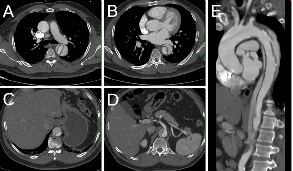
Figure 1.
A. Axial CT. Note inflated intra-aortic balloon (red arrows). in descending thoracic aorta B. IABP inflated in descending thoracic aorta (red arrow). C. Sagittal CT showing IABP inflated in descending thoracic aorta (red arrow).

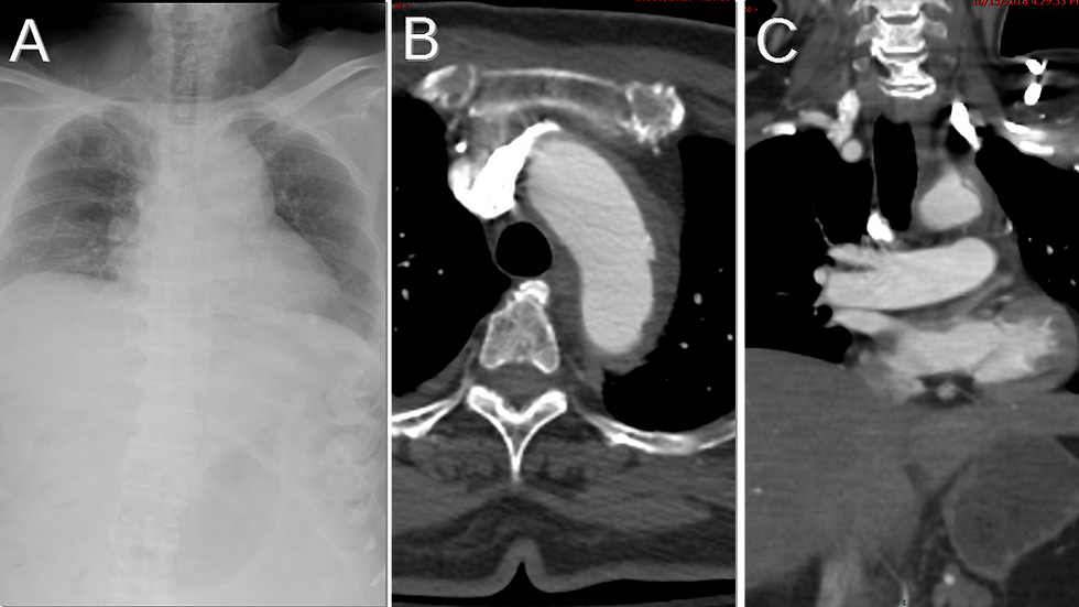
Figure 2. Frontal chest X-ray showing the intra-aortic balloon inflated in the descending aorta (red arrows). Note the radiopaque marker at the tip (yellow arrow).

Figure 3. Diagram demonstrating intra-aortic balloon inflated in diastole and deflated in systole. Image from https://www.ncbi.nlm.nih.gov/pmc/articles/PMC3177429/ (Ref.5)
Figure 4. Video of IABP function.
Discussion:
The intra-aortic balloon pump (IABP) is a cardiac assist device. It is used to stabilize patients with hypoperfusion due to acute congestive heart failure, acute mitral regurgitation, myocardial infarction, or low cardiac output after coronary artery bypass grafting surgery (1). It can also be used as prophylaxis in high risk percutaneous coronary intervention (1). The IABP has a polyethylene or polyurethane balloon at the distal tip of a large bore catheter connected to a console that inflates the balloon with helium (2). The catheter is inserted into the aorta through the femoral artery and the balloon sits in the aorta, 2.5 cm from the left subclavian artery (3).
During diastole, the balloon appears radiolucent structure in the descending thoracic aorta (Figs. 1,2). The tip is marked by a linear metallic density on imaging (Fig. 2) (3). The intra-aortic balloon uses counterpulsation to inflate and deflate. It inflates in diastole during aortic valve closure, which increases blood flow to the coronary arteries through retrograde flow (4). It deflates at onset of ventricular systole to increase the cardiac output by decreasing afterload (Fig. 4) (1,4). Overall, the IABP reduces left ventricular wall stress and reduces myocardial oxygen demand while improving stroke volume (4). Contraindications to IABP include uncontrolled sepsis, uncontrolled bleeding diathesis, moderate to severe aortic regurgitation, aortic aneurysm, aortic dissection, or severe peripheral artery disease that has not been pretreated with stenting (1,4). Balloon entrapment and rupture can occur, resulting in gas embolus (4). Improper positioning of the IABP can cause cerebral or renal compromise (4).

References:
Khan, Tahir M., and Abdul H. Siddiqui. “Intra-Aortic Balloon Pump (IABP).” StatPearls, StatPearls Publishing, 2020. PubMed, http://www.ncbi.nlm.nih.gov/books/NBK542233/
Francolini, I., and A. Piozzi. “12 - Antimicrobial Polyurethanes for Intravascular Medical Devices.” Advances in Polyurethane Biomaterials, edited by Stuart L. Cooper and Jianjun Guan, Woodhead Publishing, 2016, pp. 349–85. ScienceDirect, doi:10.1016/B978-0-08-100614-6.00012-3
Dipoce, J., et al. “Radiology of Cardiac Devices and Their Complications.” The British Journal of Radiology, vol. 88, no. 1046, Feb. 2015. PubMed Central, doi:10.1259/bjr.20140540
Krishna, Murli, and Kai Zacharowski. “Principles of Intra-Aortic Balloon Pump Counterpulsation.” Continuing Education in Anaesthesia Critical Care & Pain, vol. 9, no. 1, Feb. 2009, pp. 24–28. academic.oup.com, doi:10.1093/bjaceaccp/mkn051
Ginat D, Massey HT, Bhatt S, Dogra VS. Imaging of mechanical cardiac assist devices. J Clin Imaging Sci. 2011;1:21. doi:10.4103/2156-7514.80373

Amara Ahmed is a medical student at the Florida State University College of Medicine. She serves on the executive board of the American Medical Women’s Association and Humanities and Medicine. She is also an editor of HEAL: Humanism Evolving through Arts and Literature, a creative arts journal at the medical school. Prior to attending medical school, she graduated summa cum laude from the Honors Medical Scholars program at Florida State University where she completed her undergraduate studies in exercise physiology, biology, and chemistry. In her free time, she enjoys reading, writing, and spending time with family and friends.
Follow Amara Ahmed on Twitter @Amara_S98

Kevin M. Rice, MD is the president of Global Radiology CME
Dr. Rice is a radiologist with Renaissance Imaging Medical Associates and is currently the Vice Chief of Staff at Valley Presbyterian Hospital in Los Angeles, California. Dr. Rice has made several media appearances as part of his ongoing commitment to public education. Dr. Rice's passion for state of the art radiology and teaching includes acting as a guest lecturer at UCLA. In 2015, Dr. Rice and Natalie Rice founded Global Radiology CME to provide innovative radiology education at exciting international destinations, with the world's foremost authorities in their field. In 2016, Dr. Rice was nominated and became a semifinalist for a "Minnie" Award for the Most Effective Radiology Educator.
Follow Dr. Rice on Twitter @KevinRiceMD


Comments