


Anterior Shoulder Dislocation and Hill-Sachs Defect
Trauma. Left shoulder pain. Diagnosis? • Radiology Case of the Week
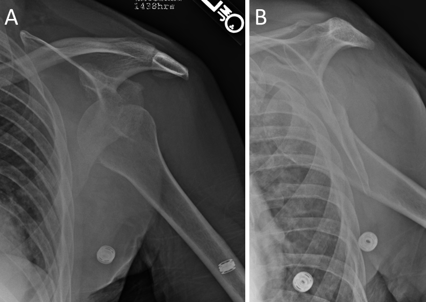
Figure 1. Left shoulder radiographs. Frontal (A) and scapular Y view (B). Trauma. Left shoulder pain. Diagnosis? Eponym?

Figure 2. Left shoulder radiographs. Frontal (A) and scapular Y view (B). Anterior dislocation with the humeral head anterior to the glenoid (yellow arrows). Also note the fracture of the superolateral humeral head (red arrows) indicating a Hill-Sachs defect.

Figure 3. Left shoulder frontal radiograph. Anterior dislocation has been reduced so the humeral head is now within the glenoid (yellow arrows). Also note the impaction fracture of the superolateral humeral head (red arrows) indicating a Hill-Sachs Defect.
Epidemiology
A Hill-Sachs defect is an impaction fracture of the posterolateral humeral head, first described by Hill and Sachs in 1940 [1]. It is very common after anterior glenohumeral dislocation: it is present in about 7 in 10 patients after a first dislocation and in more than 8 in 10 after recurrent dislocation [2,3]. In published series, most patients are young men [4].
Clinical Findings
The presence of the Hill-Sachs defect indicates that a complete anterior glenohumeral dislocation has occurred, even if the joint has since been reduced [4,5]. A large humeral head defect can engage the anterior glenoid rim, causing apprehension, popping or catching, and recurrent instability [6].
Pathology During anterior dislocation, the posterolateral humeral head is driven against the anteroinferior glenoid rim, producing a compression (impaction) fracture of the articular surface and underlying subchondral bone. The larger the humeral head defect, and the greater the associated glenoid bone loss, the more likely the defect is to engage the glenoid rim and cause recurrent dislocation [7,8].
Radiographic Features
Radiographs are the first-line study. The lesion appears as a groove-like impaction defect of the superolateral humeral head and is best seen on the AP view in internal rotation. The Stryker notch view demonstrated the defect in 25 of 27 shoulders in one study, compared with 7 of 27 on a standard frontal view, so additional views are recommended when the lesion is suspected. CT best defines the size of the defect.
In this case, Figure 2 shows anterior dislocation of the humeral head, which is displaced from the glenoid fossa and the defect of the superolateral humeral head. Post-reduction images (Fig. 3) often show the Hill-Sachs defect to better advantage.
Treatment and Prognosis
Management depends on the size of the humeral head defect and whether it engages the glenoid rim. Engaging Hill-Sachs lesions are an important cause of failure of arthroscopic Bankart repair and should be identified preoperatively, Rowe recommended surgical management of severe lesions [5,6]. One option is remplissage, in which the infraspinatus tendon and posterior capsule are fixed into the defect. Because prevalence rises with recurrent dislocation, early stabilization after a first dislocation has been suggested when the lesion is present.
Conclusion
A Hill-Sachs lesion is an impaction fracture of the posterolateral humeral head that marks a previous anterior glenohumeral dislocation. It is common, especially after recurrent dislocation. Radiographs usually demonstrate it, CT defines its size, and large engaging lesions may require surgical treatment.

References:
Ors A, Colak T, Buyukmurat N, Erdemir C, Kibar B. Relationship between Hill-Sachs lesions and morphometry of glenoid, acromion and coracoid process. North Clin Istanb. 2025;12(6):667-673. Published 2025 Dec 10. DOI: 10.14744/nci.2025.51447
Calandra JJ, Baker CL, Uribe J. The incidence of Hill-Sachs lesions in initial anterior shoulder dislocations. Arthroscopy. 1989;5(4):254-257. DOI: 10.1016/0749-8063(89)90138-2
Rutgers C, Verweij LPE, Priester-Vink S, van Deurzen DFP, Maas M, van den Bekerom MPJ. Recurrence in traumatic anterior shoulder dislocations increases the prevalence of Hill-Sachs and Bankart lesions: a systematic review and meta-analysis. Knee Surg Sports Traumatol Arthrosc. 2022;30(6):2130-2140. DOI: 10.1007/s00167-021-06847-7
Rozing PM, de Bakker HM, Obermann WR. Radiographic views in recurrent anterior shoulder dislocation. Comparison of six methods for identification of typical lesions. Acta Orthop Scand. 1986;57(4):328-330. DOI: 10.3109/17453678608994403
Rowe CR, Patel D, Southmayd WW. The Bankart procedure: a long-term end-result study. J Bone Joint Surg Am. 1978;60(1):1-16. https://pubmed.ncbi.nlm.nih.gov/624747/
Burkhart SS, De Beer JF. Traumatic glenohumeral bone defects and their relationship to failure of arthroscopic Bankart repairs: significance of the inverted-pear glenoid and the humeral engaging Hill-Sachs lesion. Arthroscopy. 2000;16(7):677-694. DOI: 10.1053/jars.2000.17715
Yamamoto N, Itoi E, Abe H, et al. Contact between the glenoid and the humeral head in abduction, external rotation, and horizontal extension: a new concept of glenoid track. J Shoulder Elbow Surg. 2007;16(5):649-656. DOI: 10.1016/j.jse.2006.12.012
Di Giacomo G, Itoi E, Burkhart SS. Evolving concept of bipolar bone loss and the Hill Sachs lesion: from "engaging/non-engaging" lesion to "on-track/off-track" lesion. Arthroscopy. 2014;30(1):90-98. DOI: 10.1016/j.arthro.2013.10.004

Umaiza Ihsan, MD is a medical graduate of King Edward Medical University, Pakistan. She completed her internship at Mayo Hospital, Lahore, including a three month rotation in Radiology, which further strengthened her interest in diagnostic imaging. She has gained additional clinical exposure in Radiology through observerships at Mon Health Morgantown and the University of Maryland, Baltimore. She has a strong interest in Radiology, particularly diagnostic imaging and its role in clinical decision making, and is actively interested in clinical research, with experience in systematic reviews and medical research. All posts by Umaiza Ihsan, MD

Kevin M. Rice, MD is the president of Global Radiology CME and is a radiologist with Cape Radiology Group. He has held several leadership positions including Board Member and Chief of Staff at Valley Presbyterian Hospital in Los Angeles, California. Dr. Rice has made several media appearances as part of his ongoing commitment to public education. Dr. Rice's passion for state of the art radiology and teaching includes acting as a guest lecturer at UCLA. In 2015, Dr. Rice and Natalie Rice founded Global Radiology CME to provide innovative radiology education at exciting international destinations, with the world's foremost authorities in their field. In 2016, Dr. Rice was nominated and became a semifinalist for a "Minnie" Award for the Most Effective Radiology Educator. He was once again a semifinalist for a "Minnie" for 2021's Most Effective Radiology Educator by AuntMinnie.com. He has continued to teach by mentoring medical students interested in radiology. Everyone who he has mentored has been accepted into top programs across the country including Harvard, UC San Diego, Northwestern, Vanderbilt, and Thomas Jefferson.
Follow Dr. Rice on Twitter @KevinRiceMD


Comments