


Kommerell Diverticulum
A 58-year-old male, asymptomatic. Identify the indicated structures and state the potential life-threatening complication. • Radiology Case of the Week
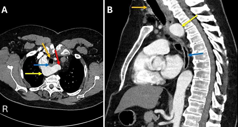
Figure 1. Name the anatomy and potential life-threatening complication.
Figure 2. A. Axial CT of the chest. The right-sided aortic arch lies to the right of the air-filled trachea (orange arrow), with the esophagus (blue arrow) situated immediately posterior to the trachea. An aberrant left subclavian artery (red arrow) arises from a focal, aneurysmal outpouching of the proximal descending aorta, the Kommerell diverticulum (yellow arrow). B. Sagittal CT of the chest. The Kommerell diverticulum (yellow arrow) is seen posterior to the esophagus (blue arrow). The air-filled trachea (orange arrow) is marked.
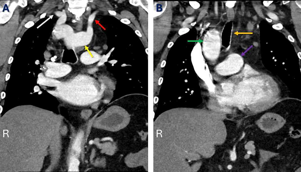
Figure 3.
A. Coronal CT of the chest, posterior projection. The right-sided aortic arch gives origin to an aberrant left subclavian artery (red arrow) that arises from the Kommerell diverticulum (yellow arrow). B. Coronal CT of the chest, anterior projection. The right-sided ascending aorta (green arrow) is seen on the right side of the air-filled trachea (orange arrow). The main pulmonary artery (purple arrow) is also well seen.
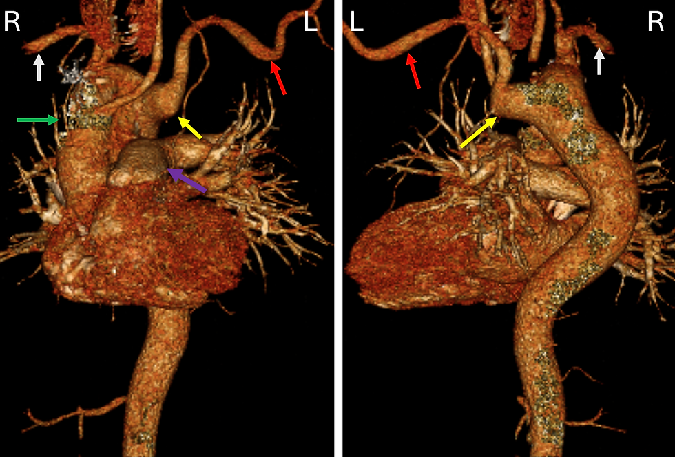
Figure 4.
A. Volume-rendered CT, anterior view. The ascending aorta (green arrow) gives rise to the right subclavian artery (white arrow) as its first branch; the right-sided aortic arch continues to the origin of the aberrant left subclavian artery (red arrow) arising from the Kommerell diverticulum (yellow arrow), with the main pulmonary artery (purple arrow) seen anteriorly. B. Volume-rendered CT, posterior view. The Kommerell diverticulum (yellow arrow) is again seen giving rise to the aberrant left subclavian artery (red arrow) from the right-sided aortic arch, which also gives rise to the right subclavian artery (white arrow).
Epidemiology
Kommerell diverticulum is a rare congenital anomaly of the aortic arch that is associated with an aberrant subclavian artery in the majority of reported cases, with most series describing an association in roughly 60–80% of patients [1]. Because an aberrant right subclavian artery is itself uncommon, occurring in approximately 0.5–2% of the general population, and an aberrant left subclavian artery is markedly rarer still, occurring in only about 0.04–0.4% of the population, Kommerell diverticulum with a right-sided aortic arch and aberrant left subclavian artery, as in the present case, represents one of the least frequently encountered configurations of this anomaly [1].
Clinical Findings
Kommerell diverticulum is frequently discovered incidentally when cross-sectional imaging is performed for an unrelated indication, as illustrated by the asymptomatic 58-year-old man described [2]. When symptoms do occur, they most often relate to mechanical compression of the adjacent trachea and esophagus by the diverticulum and its associated vascular ring, presenting as dysphagia and dyspnea [3]. Less commonly, patients may present with chest pain or with embolic phenomena arising from mural thrombus within the diverticulum. The most feared complications, though uncommon, are aortic dissection and frank rupture of the diverticulum.
Pathology Embryologically, the Kommerell diverticulum represents the persistent remnant of the dorsal portion of the fourth aortic arch [4]. In the setting of a right-sided arch specifically, it reflects persistence of the right fourth aortic arch together with involution of the left fourth arch, which normally forms the definitive arch in the more common left-arch configuration [5,6]. Histologic examination of resected diverticular tissue has demonstrated cystic medial necrosis within the wall of the diverticulum in a substantial proportion of cases, a finding that likely explains the propensity of this lesion toward dissection and rupture [7] which can be life-threatening.
Classification Three principal patterns of aortic arch diverticulum have traditionally been described in the literature: a left-sided aortic arch with an aberrant right subclavian artery, which is the most common configuration; a right-sided aortic arch with an aberrant left subclavian artery, the pattern demonstrated in the present case; and, less commonly, a diverticulum situated at the aortoductal junction. When a right-sided aortic arch is present, further sub-classification follows the Edwards system, in which type I demonstrates mirror-image branching without a retroesophageal component, type II, the pattern seen here, consists of an aberrant left subclavian artery arising from a Kommerell diverticulum, and type III, the rarest variant, consists of an isolated left subclavian artery that communicates instead with the pulmonary artery via a ductal remnant [4].
Radiographic Features
The aberrant subclavian artery can be seen to arise from a focal, saccular dilatation of the proximal descending thoracic aorta (Figs. 1-4). The diverticulum itself is conventionally measured in cross-section at the level of the origin of the aberrant subclavian artery, and in a right-sided arch such as this one, the arch and its proximal descending continuation characteristically lie to the right of the trachea rather than to the left (Fig. 1). Careful attention should also be paid to the caliber of the adjacent trachea and esophagus, since both structures may be compressed by the diverticulum and its associated ligamentous or ductal remnants.
Treatment and Prognosis
Small, asymptomatic diverticula can generally be managed with periodic imaging surveillance alone, since growth tends to be slow, on the order of roughly 1.5 mm per year in reported series. Most studies favor surgical or endovascular repair once the diverticulum orifice exceeds approximately 30 mm, once the adjacent descending aorta exceeds approximately 50 mm, or once the patient becomes symptomatic from airway or esophageal compression [7,8]. In one large surgical series, at least one death from aortic rupture has been reported in a nonoperatively managed patient whose diverticulum measured only 3.4 cm, a reminder that even moderately sized lesions warrant close follow-up.
Conclusion
Kommerell diverticulum is an uncommon congenital anomaly of the aortic arch that is most often identified incidentally in asymptomatic adults. Because the lesion is closely associated with an aberrant subclavian artery and carries a recognized risk of dissection and rupture, accurate characterization is essential once it is discovered. The three-dimensional reconstructions such as shown in Fig.4 are particularly valuable for delineating the overall arch anatomy and for planning any subsequent operative or endovascular approach [9].

References:
Irshad S, Adrejiya P, Abubaker M, Whitaker J. Kommerell's Diverticulum Masquerading in a Right Aortic Arch: A Vascular Surprise. Methodist Debakey Cardiovasc J. 2025;21(1):81-83. Published 2025 Aug 20. DOI: 10.14797/mdcvj.1641
Erben Y, Brownstein AJ, Velasquez CA, Li Y, Rizzo JA, Mojibian H, Tanweer M, Zafar MA, Ziganshin BA, Elefteriades JA. Natural history and management of Kommerell's diverticulum in a single tertiary referral center. J Vasc Surg. 2020;71(6):2004-2011. DOI: 10.1016/j.jvs.2019.08.260.
Vinnakota A, Idrees JJ, Rosinski BF, et al. Outcomes of Repair of Kommerell Diverticulum. Ann Thorac Surg. 2019;108(6):1745-1750. DOI: 10.1016/j.athoracsur.2019.04.122
Kanwal D, Khalil S, Attia K, Fam M, Arakkal M. Kommerell's diverticulum with right-sided aortic arch and anomalous origin of neck vessels: Uncommon imaging finding in neonate with cyanosis. BJR Case Rep. 2021;7(4):20200173. Published 2021 Apr 30. DOI: 10.1259/bjrcr.20200173
Summa CH, DeMaioribus CA, Swink D, et al. Hybrid repair of Kommerell's diverticulum with aberrant subclavian artery. J Vasc Surg Cases Innov Tech. 2026;12(2):102129. Published 2026 Jan 5. DOI: 10.1016/j.jvscit.2026.102129
Mubarak MY, Kamarul AT, Noordini MD. Right-sided Aortic Arch with Aberrant Left Subclavian Artery from Kommerell's Diverticulum. Iran J Radiol. 2011;8(2):103-106. https://pmc.ncbi.nlm.nih.gov/articles/PMC3522316/
Tanaka A, Milner R, Ota T. Kommerell's diverticulum in the current era: a comprehensive review. Gen Thorac Cardiovasc Surg. 2015;63(5):245-259. DOI: 10.1007/s11748-015-0521-3
Silva AF, Dos Santos JA. Aortic arch anomaly in an adult patient: a case of right aortic arch with aberrant left subclavian artery and Kommerell's diverticulum. Radiol Bras. 2016;49(4):274-275. DOI: 10.1590/0100-3984.2015.0087
Osawa H, Shinohara D, Orii K, et al. Right Aortic Arch and Kommerell's Diverticulum Repaired without Reconstruction of Aberrant Left Subclavian Artery. Case Rep Vasc Med. 2013;2013:840804. DOI: 10.1155/2013/840804

Umaiza Ihsan, MD is a medical graduate of King Edward Medical University, Pakistan. She completed her internship at Mayo Hospital, Lahore, including a three month rotation in Radiology, which further strengthened her interest in diagnostic imaging. She has gained additional clinical exposure in Radiology through observerships at Mon Health Morgantown and the University of Maryland, Baltimore. She has a strong interest in Radiology, particularly diagnostic imaging and its role in clinical decision making, and is actively interested in clinical research, with experience in systematic reviews and medical research.

Kevin M. Rice, MD is the president of Global Radiology CME and is a radiologist with Cape Radiology Group. He has held several leadership positions including Board Member and Chief of Staff at Valley Presbyterian Hospital in Los Angeles, California. Dr. Rice has made several media appearances as part of his ongoing commitment to public education. Dr. Rice's passion for state of the art radiology and teaching includes acting as a guest lecturer at UCLA. In 2015, Dr. Rice and Natalie Rice founded Global Radiology CME to provide innovative radiology education at exciting international destinations, with the world's foremost authorities in their field. In 2016, Dr. Rice was nominated and became a semifinalist for a "Minnie" Award for the Most Effective Radiology Educator. He was once again a semifinalist for a "Minnie" for 2021's Most Effective Radiology Educator by AuntMinnie.com. He has continued to teach by mentoring medical students interested in radiology. Everyone who he has mentored has been accepted into top programs across the country including Harvard, UC San Diego, Northwestern, Vanderbilt, and Thomas Jefferson.
Follow Dr. Rice on Twitter @KevinRiceMD


Comments